



CX 2026 Registration
CX Executive Board
CX 2026 Programme
CX 2026 – The 48th Charing Cross Symposium
The Charing Cross (CX) Symposium continues to set the standard in vascular and endovascular education, shaping the future of practice worldwide. CX 2026 marked the final chapter of our current three-year cycle, designed to spark debate on the biggest controversies in the field. Our distinguished faculty of global experts critically examined the evidence, exchanged perspectives, and worked towards building consensus on the issues that matter most.
Consensus was the defining theme for CX 2026. We welcomed an international community of specialists, educators, and innovators to tackle the pressing challenges in vascular and endovascular medicine—challenges that directly impact patient outcomes and clinical decision-making.
In 2024, CX found a new home at ExCeL London, unlocking fresh opportunities for immersive, hands-on learning. The expanded workshop programme has gone from strength to strength, offering delegates practical skills alongside groundbreaking evidence and innovation. In 2026, we returned once again to London’s premier conference and exhibition venue to deliver an agenda packed with cutting-edge science, expert debate, and future-shaping education.
Stay connected—sign up for the CX Newsletter and be the first to hear the latest updates.
CX Co-Chairs


Latest CX news
CT is the best imaging modality for deep vein thrombosis
Gerard O’Sullivan, Galway, Ireland, spoke in the Venous Challenges sessions on Tuesday 9 April about imaging for iliac deep vein thrombosis and the best modalities to use. He told delegates that computed tomography pulmonary angiography (CTPA) and CT venography are his preferred modalities.
I do a lot of acute iliofemoral deep venous work, and I do not think there is one modality that covers it all. If I had lots of MRI scanners and a lot of time I would use MRI much more, but I do not. When you are doing iliofemoral deep venous work for thrombosis, what kills the patient is pulmonary embolism and right ventricular dilatation. In my opinion, you need to evaluate the right ventricle and the pulmonary arteries when you are evaluating this patient. You can do that by a variety of techniques, but for me CTPA is the quickest,” he said.
Comparing CTPA and CT venography vs. MR venography, O’Sullivan noted that CT is quicker but involves the use of radiation. “It is less elegant but you can see more the inside of vessels,” he stated. “We looked at our data and, despite previous works suggesting that one-third of patients had positive CTPA with deep vein thrombosis, we found that 79% of our patients had had a pulmonary embolism by the time they presented for iliofemoral deep venous acute treatment.”
He explained that what physicians should be looking for is not pulmonary embolism, but right ventricular dilatation. O’Sullivan said that the pulmonary arteries should be assessed in several ways: isotope plus direct venography, isotope or CTPA plus ultrasound of the legs, CTPA plus CT venography, MR pulmonary angiogram plus peripheral MR venography, or echocardiography plus peripheral MR venography.
“In practical terms, for me CTPA plus CT venography is the better method as I can do it quickly and have all the information I need. I also use ultrasound to assess the popliteal vein as the further down you go more difficult it is to interpret CTPA,” he said.
He explained that in Galway he performs indirect CV venography with standard peripheral intravenous injects at same sitting at CTPA, 20G cannula in wrist or elbow, 150cc iodinated contrast, image at 150s, and 5mm cuts from diaphragm to mid-calf. “The pictures are not comparable to CT angiography but with experience are easily adequate for diagnosis.”
O’Sullivan concluded by saying that CT venography and MR venography are essential in addition to ultrasound in preoperative assessment of iliac venous thrombosis. “We feel limited ultrasound with CTPA and indirect CT venography offers the most rapid diagnostic combination method to assess pulmonary embolus, right ventricular dilatation, inferior vena cava thrombus, acute vs. chronic disease above and below the groin and extra-vascular lesions eg tumour. But other physicians use other methods equally well or better.”
Renal denervation should not be part of routine clinical practice, delegates say
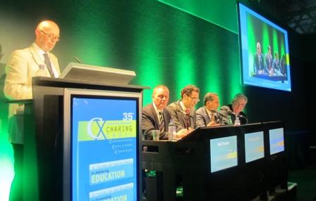
At today’s CX Renal Denervation Session Great Debate, delegates voted 3:1 against the motion that renal denervation should be used routinely to treat hypertension. As all of the speakers, regardless of which side of the debate they were, agreed that renal denervation should only be used to treat patients with resistant hypertension, the real focus of the debate centred on what was meant by the word “routine”.
Mel Lobo (London, UK), who was supporting the motion that renal denervation should be part of routine clinical practice to treat hypertension, claimed that the definition of routine was a “key issue” in the debate. He explained that patients who were suitable candidates for the intervention belonged to a highly selective group of patients, who had true resistant hypertension (ie. non-concordance with medication and other causes of resistant hypertension had been ruled out) and who would be managed by hypertension specialists rather than by primary care physicians. Lobo added that, in his view, renal denervation should become “step five” of the UK National Institute for Health and Clinical Excellence (NICE) pathway for the management of hypertension, commenting: “We do not have any randomised controlled trial data for step four [the use of spironolactone; only observational data are available], so why not have renal denervation as step five? Believe me when I say adding in a sixth or seventh antihypertensive drug is not going to work.”
Felix Mahfoud (Homburg, Germany), who was Lobo’s co-proponent of the motion, agreed that the routine use of renal denervation referred to it being used only in the “very specialised” group of patients with true resistant hypertension. He said: “Is it really ethical not to offer patients a modality that could benefit them?”
Speaking against the motion, Mark Caulfield (London, UK) disputed that “routine” could refer to an intervention being used in a highly selected group of patients. Caulfield commented: “Routine use means everybody; it implies that the intervention should probably be used in a large number of patients. Renal denervation is not ready to be used in routine clinical practice because there is not evidence base to support it being used in this way.” He added that the NICE consensus document on renal denervation, of which he was a co-author, was specifically drawn up because there were concerns that the doctors would use the intervention for all patients with hypertension rather than just those with true resistant hypertension.
Also speaking against the motion was Bryan Williams (London, UK), who said he was doubtful about the data from renal denervation studies. He commented: “What is critical is how many patients in these studies were actually taking their antihypertensive medication? If the drop in blood pressure observed in these studies is really a result of renal denervation on top of antihypertensive medication, then that is impressive. But if it is actually a result of patients starting to take their drugs [because they are now in a clinical trial], then it is not impressive.”
Lobo responded by saying there had to be element of trust and that “We have to believe our patients when they say they take their medication. We are not able to follow them home and check up on them.”
At the end of the debate, 3:1 delegates voted to support Caulfield and Williams that renal denervation should not be part of routine clinical practice for managing hypertension. Summing up the data, the chair of the session Neil Poulter (London, UK) said he hoped that by the time of CX36 (12–15 April 2014), more data on renal denervation would be available and the issue could be further discussed.
Endovascular management of ascending aortic pathology

By Ralf Kolvenbach
The incidence of thoracic aortic aneurysms is estimated to be as high as six cases per 100,000 person-years, and replacement of the ascending aorta accounts for the majority of cardiothoracic aortic procedures.
Aneurysms and dissections of the ascending aorta are still mainly treated operatively with cardiopulmonary bypass. Ascending aortic aneurysms with normal sinuses and aortic annulus require only replacement of the ascending aorta from the sinotubular ridge to the origin of the innominate artery with a Dacron tube graft.
Due to the significant morbidity and mortality of these procedures high-risk patients are not considered for cardiac surgery. There are a number of anecdotal reports describing endovascular stent grafting of various pathologies including deployment of a fenestrated stent graft in a case of an ascending aortic rupture.
Endovascular treatment of the ascending aorta is particularly challenging because of the anatomical features of this aortic segment. Acute and chronic Type A dissections can be treated with a tubular endograft when the aortic valve and the coronary arteries are not involved. An analysis in our clinic of 30 consecutive patients admitted with ascending aortic pathology showed that in 75% ascending aneurysms had a conical shape without a proximal landing zone. Only in a few cases the ascending aorta had a tubular configuration with a long proximal landing zone of at least 2cm. This makes over-sizing and an optimal graft configuration particularly important. Only patients without connective tissue disorders, clinically relevant aortic incompetence, stenosis or concomitant coronary artery disease can be considered for an endovascular procedure.
Material and methods
So far only patients with ascending aortic pathology who were considered unfit for open surgery were treated with an endograft. We also excluded patients with malperfusion or any preoperative unstable clinical condition after Type A dissection. Also excluded were patients with severe aortic valvular disease including aortic valvular incompetence as a consequence of an acute Type A dissection, coronary artery disease requiring surgery, and any kind of connective tissue disorder. Included in our ongoing study are patients with intramural hematoma, floating thrombus after chronic Type A dissection and penetrating aortic ulcers. Also included are aneurysm patients without aortic valve incompetence or significant dilatation of the aortic annulus requiring composite graft replacement of the aortic valve and the ascending aorta.
Patients with asymptomatic penetrating aortic ulcers (PAU) were initially managed conservatively. If symptoms did not resolve, or patients continued complaining about chest pain, endovascular exclusion was discussed with the patient.
Threshold for aneurysm patients is an aortic diameter of 6cm or larger and an adequate landing zone proximal to the coronary arteries. Patients were excluded if they were good candidates for open surgery. In cases without a good landing zone an aortic banding procedure using a mini sternotomy was performed prior to stent graft deployment as originally described by the Zurich group of Lachat.
The length of the stent graft was depending on the length of the outer curve of the ascending aorta which was regularly longer than the distance determined by center line measurement.
All operations were performed under general anesthesia. In addition to our monitoring protocol for TEVAR transoesophageal ultrasound was performed to control cardiac and valvular function. Cardiac arrest was induced with adenosine administration when required. A temporary ventricular pacemaker was placed through the jugular vein.
After road map angiography selective coronary angiography was performed to outline the origin of the coronary arteries.
An ultra stiff guide wire (Lunderquist, Cook) was placed into the left ventricle after passage of the aortic valve with a vertebral catheter over a 0.035 guide wire. The endograft (Cook custom made ascending endograft) was carefully placed across the aortic valve into the left ventricle. The kind of graft used depended on the size required.
Tracking of the stiff wire all the time during the procedure was essential to avoid ventricular perforation. At the end of the procedure ventriculography was performed to rule out any damage to the left ventricle or the valve apparatus. In case of any ECG changes coronary angiography was added before removing the catheter and sheaths. Before discharge a contrast enhanced angio CT was performed as well as cardiac ultrasound examination.
Ascending aortic dilatation may be caused by intrinsic pathology of the aortic wall, or hemodynamic factors caused by a stenotic aortic valve: High velocity and turbulent flow downstream of the stenosis place mechanical stress on the aortic wall.
The thin wall of the ascending aorta does not permit aggressive over dilatation. The graft was deployed in most cases without any additional balloon dilatation. The walls of the aortic sinus (sinus of Valsalva) are considerably thinner than the wall of the aorta. Hooks or bear springs should therefore be deployed in a safe distance from the sinus and the origin of the coronary arteries to avoid erosion and perforation. One of our first patients suffered from a stroke postoperatively.
Calcifications and thrombus lining of the ascending aorta and aortic arch were the main parameters for increased stroke risk
Access can be a problem since in most aneurysm cases a 24F access sheath was required. In two cases with sever calcification of the iliac vessels we had to use the left carotid artery as an access which proved to be uneventful. Alternatively using a small thoracotomy the endograft can be deployed through the apex of the heart.
There is so far only one dedicated graft for the ascending aorta (Cook). Since most aneurysms have conical shape a stent graft designed for Type a dissection cases is not necessarily suitable for aneurysm patients. So far the anchoring zone of a few millimeters only, requires fixation with hooks, significant over sizing and coil deployment into the sac of the aneurysm if necessary. Banding of the ascending aorta is an option to increase the landing zone in aneurysm patients with conical aneurysm morphology.
The aortic impulse which is directly proportional to the mean blood pressure, the cross sectional area of the aorta and the duration during systole varies inversely with the distance from the aortic valve. In stent grafting of the aortic arch and the descending aorta high aortic impulses can cause significant pulsate motion of the arch and inadvertent movement of the stent graft. In the future more active fixation with a stapler would probably permit safer deployment, less Type I leaks and better long term performance. Especially when treating more advanced stages of aortic pathology a valve bearing conduit with fenestrations for the coronary arteries will be necessary.
Before a routine use can be advocated outside of studies several issues must be addressed. There should be dedicated grafts for ascending aneurysms and for acute dissections like the one available so far. Precise deployment of the graft adjacent to the coronary arteries is essential in aneurysm patients. In most cases there is a landing zone of a few millimeters only, deployment too distal from the sinotubular junction results in kinking of the graft and subsequently a type I leak.
We can conclude that stent grafting of the ascending aorta is technically feasible but should be reserved to selected high risk patients only, preferably in centers where vascular specialists cooperate closely with interventional cardiologists. Cardiac surgery with cardiopulmonary bypass and if necessary deep hypothermia is still the gold standard when treating ascending aortic aneurysms though still associated with significant morbidity and mortality. Stent graft exclusion of more advanced and complex ascending aortic aneurysms should be reserved for high risk patients only in centers with the necessary skills to perform transvalvular cardiac procedures. This may change in the future when more dedicated grafts become available. The future will and has to show valve bearing endovascular conduits which will permit minimal invasive treatment of most aortic aneurysms. The grafts available so far already permit emergency endovascular treatment of type A dissections. Yet in aneurysm patients compromises are still necessary.