Controversies Challenges Consensus
}
CX News2025-01-29T14:37:45+00:00

CX NEWS

Mid-term results for endovenous ablation support shift to office-based vein practice

Setting up an office-based vein practice is becoming extremely popular as the treatment of varicose veins is undergoing a revolution. Many physicians feel that it is becoming less and less acceptable for varicose veins to be operated on by using open surgical techniques. 

Three-year results from a recently published prospective, multicentre trial using the VNUS ClosureFast catheter (Covidien) have demonstrated clinical efficacy and improvement in quality of life for patients treated for varicose veins.  Also, laser options are expanding with a new laser system, VenaCure (Angiodynamics) 1470nm laser, being launched at CX33. Recent randomised trials comparing the two modalities with short-term follow-up data have shown comparable results for efficacy.
 
Thomas Proebstle, Mannheim, Germany, presented the three-year results and exclusively told CX Daily News, “We now have solid data to support the statement that the days of cross ligation and stripping are over. We have such high success rates, good durability of results and a gain in quality of life from pre-procedure impairment compared to traditional surgery, that we need to shift to endovenous approaches of treatment.
 
“Obviously this type of treatment does not need to be performed in a theatre or hospital, which improves its cost-effectiveness. It can be performed in a modern office with excellent medical facilities, in an outpatient setting.”

Proebstle said that compared to all of the other current technologies such as laser, foam sclerotherapy and the new kid on the block, superheated steam, ClosureFast is the only one with solid mid-term follow-up. “It is the only one combining the highest efficacy with such a smooth side-effect profile. All the other technologies at the moment lack the proof that they can deliver this smooth side-effect profile, and this needs to be proved in randomised studies,” he said.
 
Shift in principle
 
Proebstle said, “After ClosureFast, the system which delivered high frequency current into the vein wall, there was a subsequent system developed which no longer delivered current to the tissue, but only heated the coil. So from a current-induced heat generated within the tissue, there was a shift in principle to heat conduction only. The catheter here is actually nothing but very expensive single use water heater. With this type of radiofrequency- powered catheter, we observe that pigmentation is a rare event and at three year follow-up, only one out of nearly 300 limbs showed persistent pigmentation.”

He explained that the three-year follow-up results were from a study of patients treated with the ClosureFast catheter at eight European centres. In the study, 295 greater saphenous veins (from 225 patients) were treated. The mean age of the patients was 50.5 + 13.5 years and 73.8% were female. The majority of the patients were CEAP C2 or C3.
 
All the patients were treated in an outpatient setting, under local anaesthesia and the protocol of heat delivery was two treatment cycles immediately below the saphenofemoral junction, and one treatment cycle subsequently thereafter. Combined procedures were allowed only at calf level, but not at the thigh. 
 
“Clinical and duplex examination took place at three days during the first week, at three months, six, 12, 24 and 36 months we had a data retrieval rate of 86–87%, if you refer to patients and limbs treated respectively after three years,” he explained.
 
Proebstle said, “These data include the first-in-man treatment up to the first 295 patients, so this paper actually includes the learning curve for this procedure.
 
Anybody will know that if you have very good results including in the learning curve period, such a technology could be seen as ‘idiot-proof’.”
 
 
Results
 
Proebstle said, “The initial ablation success rate with VNUS Closure Fast is 100%. Only five of 256 (2%) limbs showed axial reflux 36 months after the procedure and as such radiofrequency segmental thermal ablation shows a high efficacy coupled with a very moderate side effect profile. At baseline, 41.1% of limbs were free of pain. At 24 months, 99.3% (n=278) reported no pain, and between12–24 months, 96.4% (n=270) had no pain.”
 
He told delegates that at 36 months, 98.0% (n=251) reported no pain, and between 24 to 36 months, 95.7% (n=245) had no pain.
In addition to clinical efficacy, the study has shown a venous clinical severity score reduction at three-year follow-up. “Overall score improved for 256 legs available at 36 months from 3.9 ± 2.1 at baseline to 0.9 ± 1.1 at 36 months, p
 
The three-year European follow-up of endovenous radiofrequency-powered segmental thermal ablation is scheduled for publication in the Journal of Vascular Surgery in 2011. 
 
 
Questions
 
Responding to a delegate who stated that he frequently saw large veins and asked what the upper limit of the diameters of veins treated in Proebstle’s study was, Proebstle replied, “In this series, the upper limit did not reach the 2cm diameter, but in such cases of large veins you can use this system repetitively. You do not have to adhere to two cycles, you can use three or four cycles for big diameter veins. This is safe, even though it has not been published.”
 
Alun Davies, London, UK, then said “the reality is that you have just convinced us that VNUS is not as straightforward as it is being sold to us. We all use variations in how we treat a segment, so it is no longer a standard treatment. Do you agree with that?”
 
Proebstle replied, “I agree that in situations not covered in the study, in extreme situations with large diameters, there may be a necessity for dose adjustment.”
 
Davies also asked for Proebstle’s thoughts about seeing more pigmentation in severe disease. Proebstle then said that in more severe disease, there was a possibility of an increase seen in the side-effects profile, but clarified that in this study several cases with severe disease were included.
 
Lowell Kabnick, New York, USA, then asked on what basic scientific evidence the proximal two-cycle protocol was based. He said, “My research has shown that there is just a 1mm difference in coagulation zones between one and two cycles. It really has to do with wall thickness rather than the size of the vessel.”
 
However, Proebstle argued that it was to do with the size of the vessel and that this was based on “biophysics, like water running down a hill”.
 
New VenaCure 1470 laser ablation system launched at CX33
 
AngioDynamics has launched a new laser system for endovenous laser ablation of varicose veins at CX33. The new VenaCure 1470nm laser operates at a peak on the water absorption curve. The VenaCure 1470nm laser is a water-specific laser, meaning it damages the intima, or inner most layer of the vein, by targeting water as the chromophore to absorb the laser energy.
 
Since the vein structure is mostly water, this laser is able to efficiently heat the vein with little chance of collateral heating, resulting in an optimal vein ablation.
 
The VenaCure 1470nm laser is designed to work exclusively with AngioDynamics NeverTouch fibre ablation kits, maximising these two technologies and resulting in even better patient outcomes.
 
A company release says that current clinical evaluations demonstrate clinical efficacy superiority with laser versus other modalities. Post-operative pain and bruising studies combining the VenaCure 1470 laser and NeverTouch fibre are currently running in Europe. AngioDynamics said it strongly believes that the new system provides the clinician and patient with the optimal combination for the minimally invasive treatment of varicose veins. 
 
The new VenaCure 1470 laser has received the CE mark. A 510k application has been submitted to the FDA.
 
Yesterday, more than 300 delegates participated in the CX Office-based Vein Practice Course.

CX heralds era of “off-the-shelf” fenestrated grafts

Andrew Holden, Auckland, New Zealand, presented results from the preliminary clinical experience with the Ventana off-the-shelf fenestrated endovascular graft (Endologix) yesterday.
“Between 25 and 40% of infra-renal abdominal aortic aneurysms are not amenable to endovascular aneurysm repair (EVAR) using currently available devices,” Holden told delegates at an Endologix sponsored event. “The most common reason is inadequate infra-renal aortic neck anatomy (short neck or neck with adverse features such as reverse conical shape or thrombus,” he said.

Fenestrated EVAR devices (FEVAR) have been developed to treat a greater number of juxta-renal abdominal aortic aneurysm morphologies by extending the graft sealing zone into the supra-renal visceral artery segment. “Published data have shown that FEVAR is associated with lower 30-day mortality and post-procedure renal impairment when compared to open repair,” Holden stated.
 
Current FEVAR devices are customised for individual patients. The planning, construction and delivery of these devices inevitably results in procedural delays, he said. “To manage this problem, Endologix has developed the first ‘off-the-shelf’ FEVAR device with steerable fenestrations to be used in clinical practice, the Ventana fenestrated endograft.”
 
Holden explained that the Ventana device has two 3mm diameter renal fenestrations with renal sheaths pre-loaded through the fenestrations. The fenestrations can be dilated up to 8mm diameter and can be moved up to 35mm from their nominal locations. There is a 4cm deep scallop for the coeliac artery and superior mesenteric artery. The Ventana Fenestrated Proximal Extension Graft is delivered after an Endologix Powerlink bifurcated graft has been deployed with anatomic fixation at the aortic bifurcation. 
 
“There are some anatomic eligibility requirements, the most important being an ‘infra-superior mesenteric artery neck’ length of 15mm or longer,” Holden said. “A key feature of the device is that it is fully constrained while the renal arteries are being cannulated – this facilitates quick and easy cannulation of even challenging renal artery anatomies. Covered balloon expandable stents are deployed into the renal arteries to create what are essentially branch grafts. In recent cases, the Endologix Xpand covered stents have been used.”
 
To date, nine patients have been treated with the Ventana system (six in Auckland and three in Santiago, Chile). There have been no major adverse events, 100% technical success, 0% mortality and 0% conversion to open repair. 
 
“Procedural times have been very satisfactory given a ‘first in man’ trial. Seven patients have had a CT scan one month after repair – there has been 100% freedom from type I or type III endoleak and all branch arteries are patent. Interestingly, all patients have received a Ventana model where the renal fenestrations are ‘aligned’ – six have received a device with a proximal diameter of 28mm and three with a proximal diameter of 24mm,” Holden said.
 
In conclusion, he said, “the Endologix Ventana Fenestrated Graft offers an exciting ‘off-the-shelf’ FEVAR device for the management of juxta-renal abdominal aortic aneurysms. Early clinical experience has been excellent.”
 
Two designs fit 90% of patients
 
Another off-the-shelf fenestrated graft is Cook Medical’s device. “It is safe to say that an off-the-shelf design would alleviate a lot of issues,” Timothy Resch, Malmo, Sweden, told the audience during the Cook satellite symposium yesterday.

He said that Cook’s off-the-shelf fenestrated device has a three-piece modular design: the proximal component which is tubular with fenestrations; the second component which is bifurcated without a top cap; and the contralateral limb.
 
“The device is based on the fenestrated platform that we have used successfully for elective patients. It is very similar in design and incorporates a lot of those features that we know have created stability over time,” Resch said. 
 
“There is a good advantage in having only unilateral access for the renal arteries and all the small sheaths. With two designs we can fit about 90% of patients.
 
The preloaded wire through the renal fenestrations is very helpful, and the self retrieving top cap makes the procedure easier as well.”
 
Resch said the device may be available by the end of the year.

Largest hands-on office-based vein practice course in the world at CX

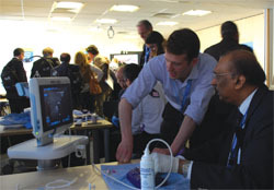
For the fourth year running, the Charing Cross Office Based Veins Course offered a vast selection of the latest therapies to treat varicose veins in an office environment. Over two sessions, 300 participants were trained on steam thermotherapy, mechano-chemo ablation, foam sclerotherapy, thread vein treatment and endovenous ablation, among other techniques.

Although the course is becoming larger it has retained its key features of small groups and one-on-one teaching with a strong emphasis on hands-on practice. CX Daily News asked Ian Franklin course director (London, UK) about the nature of the course and its evolution. “We started this course four years ago and at that time, this was the first veins course specifically aimed at looking at office-based techniques. We try to include many techniques as we possibly can and do not try to tell people what is the right or wrong way of doing things. We just offer them all the techniques and they come along and make up their own minds based on what suits their practice best,” he said. 

This year, the course has moved to more spacious surroundings. This is the “largest practical office-based vein practice course in the world,” said Franklin. “We have more space, so people are not pressured for time when learning their skills. We  also have a number of new techniques that we did not have before, so people can have the updates on the latest techniques available.” Another addition to this year’s course was the incorporation of mini symposia on essentials of setting up a successful private vein practice and also case-based discussions demonstrating the effectiveness of office-based techniques, Franklin added. 
Sherf Sholkamy, a surgeon from Cairo, Egypt, attended the course for the second year and has noticed how the course has helped him to evolve in treating thread and flare veins. “From this course, you get really good experience on how to do it and you just go home and do it,” he said. “I came a year ago to learn on the treatment of thread and flare veins and came back this year to resolve further questions, after practicing what I have learnt,” he added.  
 
As well as attendees, physicians teaching the techniques have seen the course has evolved. Ravi Singh Ranger, who was teaching venous closure for the treatment of varicose veins commented, “When I first started doing this course three years ago, these were new techniques and doctors were more interested in whether they worked or not, and if they worked how they got about doing them. What I find today is that the whole pendulum has shifted and doctors are now not questioning the validity of the techniques. They have done this before and all they want to know is what I would do if I had a really big vein, or how I manage a tortuous vein. They want to learn how to treat it perfectly.”
 
Lowell Kabnick, New York, USA, said, “Each year, the course seems to expand the amount of materials that are covered. The physicians that are passing through have more interest, more and more knowledge and so this faculty has been able to accommodate this. The hands-on models are also getting better, they are more sophisticated.This is my third year participating on this course and every year it gets much better…this is the best hands-on course and I have been to many of them.” He told CX Daily News, this is a place where physicians can come to learn about venous disease and they get it all, both from a didactic perspective and a hands-on one.”

CX News: Are the EVAR boundaries being possibly pushed too far?

Clinical practice must change today, according to the latest data from the 10-year follow-up of the UK EVAR trials and a large US M2S dataset registry. The anatomical suitability for EVAR must be respected and mandatory pre-discharge imaging should be a requirement in order to identify perioperative rupture, CX delegates heard yesterday. Remarkably, they also heard that the incidence of aortic sac enlargement after EVAR appears to be increasing, not decreasing, despite increased operator experience and improved device technology, and that this is due in part to the liberalisation of the anatomic characteristics deemed suitable for EVAR, and physicians expanding the indications for EVAR and using devices outside of the instructions for use (IFU)

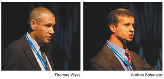
These data came both from the UK EVAR trials presented by Thomas Wyss and the large M2S study presented by Schanzer et al which was published online in Circulation immediately following presentation at CX33 yesterday. Chairman Roger Greenhalgh said the session was a historic moment, “Truly head-turning in terms of the evidence emerging,” he said.
The main finding of Thomas Wyss and colleagues, which set out to analyse graft rupture after looking at all endovascular and open repairs performed for up to 10 years in the UK EVAR trials, ­was that there were no ­ruptures after open repair. This was a surprise because ruptures after open repair have been documented in the past, as in the UK Small Aneurysm Trial.
Thomas Wyss from Bern, Switzerland, spent a year as vascular research fellow at the Charing Cross Vascular Surgery Research Group. He had access to CT scans of all the EVAR trials’ long follow-up data and conducted an analysis on graft rupture. The data were published in the Annals of Surgery in November 2010.­
The aim of the graft rupture analysis, Wyss said, was to establish the incidence of secondary rupture after abdominal aortic aneurysm correction by endovascular and open repair and to investigate factors associated with secondary rupture. In total, 848 elective endovascular repairs and 594 elective open repairs were performed in EVAR 1 and 2, he told delegates. 
The EVAR 1 trial aneurysm-related mortality converged at the six-year point, having been significantly higher for open repair at four years. In these later years of follow-up there had to be an explanation for this relative deterioration in the EVAR group at late stages. “This relative improvement of open repair is due to ruptures after EVAR,” he told delegates.
The audit showed that 27 ruptures occurred after endovascular repair – 25 in EVAR 1 and two in EVAR 2. The 27 ruptures were divided into three groups: five ruptures up to 30 days (group A), five ruptures after 30 days with no complications (group B), and 17 late ruptures (>30 days) with complications were included in group C. 
“We assume that there was a technical problem behind all of group A and the first rupture (32 days) of group B. A change of protocol, to always perform a pre-discharge CT scan/ or use of any other suitable imaging modality could have revealed problems, which could have been addressed,” Wyss said. 
There are four additional sac ruptures which occurred without complications – one failed to attend CT scan follow-up. This left three (11%) which ruptured out of the blue. The patient and clinician, Wyss said, assumed that all was well and “out of the blue” a rupture occurred. “This occurrence is fortunately rare. But, two out of these three died,” Wyss noted.
 

Cluster of known endovascular complications

The remaining 17 patients (group C) had a cluster of recognisable and known endovascular complications: sac growth, endoleaks type I and III, endoleak type II with sac growth, migration or angulation.
Wyss said that the next steps are to “implement a predischarge computed tomography scan which might reveal problems and prompt immediate treatment to reduce perioperative rupture, confirm the collection and sequence of EVAR complications which predispose to rupture, re-examine the threshold for conversion to open repair and prospectively react to high risk scenarios.”
Wyss et al were surprised to see the statistically very significant factors fed in from the audit, with p=0.0001. They concluded that these results show technology needs to improve device conformability to the aorta.
“Clinical practice must change from today. We need to respect suitability of anatomy for EVAR devices, ensure mandatory pre-discharge CT scan, encourage regular patient follow-up, and correct sac expansion and its underlying cause urgently by either endovascular methods or conversion to open repair,” Wyss said.
 

60% of EVAR cases do not meet the recommendations  

A multicentre observational study conducted in the USA has found low compliance with EVAR device guidelines, and “alarmingly high” post-EVAR aneurysm sac enlargement, raising concern for the long-term risk of aneurysm rupture. The study set out to analyse a large, multicentre, prospectively acquired dataset, representative of real world EVAR practice, containing extensive baseline and postoperative anatomic imaging data. 
Andres Schanzer, Worcester, USA, presented the results of the largest investigation to date utilising detailed pre- and post-EVAR anatomic CT imaging data to identify determinants of abdominal aortic aneurym sac enlargement post EVAR. 
He said that over 70% of elective infrarenal abdominal aortic aneurysm repair in the United States are performed using EVAR. Schanzer told delegates that baseline aortic and iliac artery anatomic characteristics are the fundamental criteria for appropriate selection of patients for endovascular aortic repair and key determinants of long-term success. “In a large, multicentre cohort, we evaluated compliance with anatomic guidelines for EVAR and the relationship between baseline aortoiliac arterial anatomy and post-EVAR abdominal aortic aneurysm sac enlargement,” he said.
The investigators identified patients with at least one pre-EVAR and at least one post-EVAR computed tomography scan from the M2S, which is an imaging database, in the period 1999–2008. 
Explaining the methodology, he said, pre-operative baseline aortic and iliac material anatomic characteristics were reviewed in order to generate a standardised set of morphologic measurements for each patient. Data relating to which specific abdominal aortic aneurysm was implanted was not available. Therefore, the morphologic measurements were compared to the most liberal and the most conservative published anatomic guidelines as stated in each manufacturer’s instructions for use. The most conservative definition was: neck length >15mm, neck diameter 10mm, neck diameter
The primary study outcome was post-EVAR abdominal aortic aneurysm sac enlargement (>5mm diameter increase).
Results 

The results of the study showed that in 10,228 patients undergoing EVAR during the study period, 59% had a maximum aneurysm diameter below the 5.5cm threshold where intervention is recommended over surveillance. Only 42% of patients had anatomic characteristics that met the most conservative definition of device instructions for use and 69% met the most liberal definition of device instructions for use. 

The five-year post-EVAR rate of abdominal aortic aneurysm sac enlargement was 41%.

Schanzer et al’s study had a mean follow up 31 ± 18 months, the mean number of follow up scans was 3.03 ± 0.9. The important findings that he discussed at CX were:


    • Only 40% of aneurysms treated with EVAR meet the recommended diameter threshold for treatment (≥5.5 cm).
    • Even applying the most liberal instructions for use definition, 30% of treated patients do not meet aortic neck criteria.
    • Anatomy deemed acceptable for EVAR has continued to liberalise and several of these factors are independently associated with aortic aneurysm sac enlargement.
    • Aortic sac enlargement post EVAR occurred in 40% of patients by five years.
    • This incidence is significantly higher than that reported in industry-sponsored trials and reports from single centres of excellence.
    • In >30% of patients demonstrating aortic sac enlargement, this was not detected until >3 years postoperatively.
    • The incidence of aortic sac enlargement after EVAR seems to be increasing, not decreasing—despite increased surgeon experience and improved device technology.
Schanzer outlined that independent predictors of aortic sac enlargement included: age >80; aortic neck diameter >28mm; aortic neck angle >60 degrees; common iliac artery diameter >20mm and presence of any endoleak on follow-up.
He clarified that some of the limitations of the study were that there was no endograft specific data, no data related to re-interventions, non-consecutive submission of patients to this dataset may introduce selection bias and limit generaliseability. Also, there were no clinical outcomes data available.
Schanzer told delegates that, “Liberalisation of the anatomic characteristics deemed suitable for EVAR has occurred and this is associated with a significant increase in the incidence of aortic sac enlargement thereby reducing durability of EVAR to protect against abdominal aortic aneurysm rupture. 
“An honest appraisal of current real world outcomes, with a prospective registry that incorporates blinded imaging review is warranted,” said Schanzer. He also went on to ask if current available commercial devices are truly appropriate for the increasing complex anatomy that they are being applied to?”
Voting results
When the audience of specialists was asked whether they were fixing aortic aneurysms less than 5.5cm in diameter, there was a nearly equal split, with 52% saying that they were fixing such aneurysms and 48% saying they were not. Greenhalgh said, “No matter what the randomised controlled trial data are, several people are breaching that knowingly. Now you have learned that you can get the same problems putting in an endograft unnecessarily and I hope you take his message to heart.” 
To the second question, “Should we intervene for sac expansion after EVAR with the defined cluster of factors associated with rupture?” an overwhelming majority of the audience voted that intervention is necessary. 

Go to Top