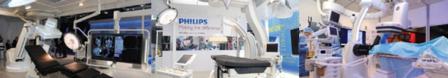
For the very first time, three full-scale sterile hybrid operating suites from GE Healthcare, Philips and Siemens were on display at CX. The importance of quality imaging as a prerequisite for improved clinical outcomes was emphasised in every section of the main programme. This went hand in hand with calls from physicians for high-quality image availability in the intraoperative setting.
Imaging is certainly at the heart of endovascular intervention, and it is now widely accepted that using the best available imaging can have a direct impact on achieving the best clinical results. With the blurring of boundaries between specialties in the endovascular arena, there was a clear need expressed by clinicians at CX for improved imaging at the intraoperative stage. Delegates at CX35 are split in nearly equal proportions along the disciplines of vascular surgery, interventional radiology and interventional cardiology.
Georg Nollert, director, Global Marketing, Siemens, told CX Daily News: “I am very happy that the focus on imaging is increasing. In the past, vascular surgeons were satisfied with inferior image quality and other interventionalists such as radiologists and cardiologists benefited from using the best available imaging. I believe that surgeons ought to have the same image quality in order to get the best results.”
In the preoperative and postoperative setting, ultrasound and other sophisticated imaging modalities such as CT or MRI are widely available. Nollert said: “Intraoperatively, however, imaging was limited to the use of C-arms (2D fluoroscopy) in the past. Therefore, one available solution was to enable the superimposition of the preoperative images with the intraoperative images. Using preoperative CT or MRI, this then creates the 3D road map that interventionalists could use for very sophisticated interventions.
Nollert told CX Daily News that in the past, the issue with cone beam CT was one of image quality, particularly with reference to contrast resolution and that Siemens has been working on improving this aspect. “We now have new algorithms to reduce metal artifacts and also better detectors in the latest family of systems, the Artis Q. Artis Zeego also has the latest technology and the latest detectors and we have increased the contrast resolution for cone beam CT substantially. It is important to bear in mind that all cone beam CT is not equal. We have new protocols that result in the quality being much superior to regular/conventional CT. However, image quality for cone beam CT depends on reconstruction algorithms, and how you deal with artifacts and distortion, and those algorithms are widely different in the market.” Kirsten Zuurmond, clinical scientist, Philips Healthcare and Koen Noordermeer, Business Development Manager, Philips Healthcare, told CX Daily News: “Philips is very concerned about the quality of intraoperative imaging. In order to reduce radiation dose and contrast values, Philips offers the possibility of fusion imaging where clinicians use the preoperative CT as a navigational map during the procedure. We also offer the flexibility of working with cone beam CT, such as in emergency situations, so that it is possible to make an accurate 3D image at the intraoperative stage. Zuurmond highlighted that clinical opinion was clearly divided on the topic with some clinicians experiencing a high degree of accuracy. She then also explained that the company was focusing on the ostia as a target in fenestrated EVAR procedures and noted that they hoped to provide the extra help that could be useful in the area. Ease of use
“Another possibility was just to use intraoperative cone beam CT, and we are getting close to conventional CT quality with this. The elegance of this solution is that the images are automatically registered to the patient and there is, on the other hand, the actual anatomy of the patient on the table that is probably not the anatomy that you have on the
He highlighted that the Artis Zeego was a flexible, robotic system that can adjust to the table and to being used by the whole team in the operating room. The system also allows for maintenance of the sterility and environment of the operating room as it keeps the ceiling free for uninterrupted laminar flow, or use of operating room lamps, for example. “With regard to 3D capability, the Artis Zeego has some absolutely unique features and can image large volumes, fast and achieve a superior image quality,” he said.
Clinicians in the session made the point that the robotic arm with the automated motion had the drawback that the user could not always predict how the system was going to move and often found that it was quite hard to anticipate the movement.
They highlighted the flexibility of positioning the c-arm Philips system within the hybrid operating room, the advantages offered by the new system Alluraclarity, in achieving significant dose reduction, without compromising on the image quality available to physicians.
3D superimposition on fluoroscopy is not yet ideal
During the Monday morning session the issue of distortion resulting from overlaying preoperative 3D images intraoperatively was discussed. A panel comprising of chairman Peter Taylor Tara Mastracci, Cleveland, USA, Krassi Ivancev and Ian Loftus, both London, UK, noted that with fusion imaging, there was no compensation for vascular distortion and that there was imprecise fusion usage for endograft positioning and cannulation of target arteries.
“One of the issues that many clinicians have with fusion imaging is its lack of responsiveness to deformation, but most believe that a fix is on the horizon,” Mastracci said. Sixty two per cent of the audience in the session also voted against the motion that fusion of preoperative datasets is ideal.
Noordermeer then added: “We see the benefit of the current technology but are of course working on improvements for the future to make it even more accurate and applicable. We are interested in is bringing solutions that are easier to use in the hybrid operating room and are working on automating workflow steps, making access easier and providing stepwise guidance for physicians.”
Gregory McIff, global director, Cardiovascular Strategic Marketing and Delphine Germain, global product manager for Hybrid OR, GE Healthcare, told CX Daily News that flexibility in the operating room, ease of use for physicians and advanced image quality were optimised to the best degree that technology allowed today in the GE systems.
In response a questions about image quality not being as good as it could be at the time of intervention, McIff noted that image quality was subjective to the user. “We know from technology that a flat panel detector gives a clearer image than an image intensifier system, so you are going to see some changes in that. But when you are dealing with aortic interventions the ability of a system to provide good imaging is perhaps, adequate. You can always have better imaging—I would love to drive a Rolls Royce, but I have to make do with my Chevrolet!”
Germain said, “We definitely emphasise that the system is easy to use. You can have the best thing in life, but it has got to be accessible, not complicated to use, too. This is an area we will keep investing in: improving the ease of use. We already have systems that allow surgeons to learn fusion imaging from tableside. With regard to superimposing preoperative imaging data within the Hybrid OR, the technique is getting complicated and it is important for us to make sure that people can use it on a routine basis.”
Both highlighted that GE focused on combining the best of the hybrid operating room with the best of the advanced imaging techniques. They drew attention to the fact that the Discovery IGS 730 was not mounted either on the wall or on the ceiling but that it could move freely in the room.
“The system confers the benefit of mobile systems that can move in the room freely so that clinicians can have access to patients, and at the same time have the ability to be brought back into imaging position and have all the advantages of a fixed system in terms of imaging such as 3D imaging and 3D fusion,” they said.