


CX 2026 Registration
CX Executive Board
CX 2026 Programme
CX 2026 – The 48th Charing Cross Symposium
The Charing Cross (CX) Symposium continues to set the standard in vascular and endovascular education, shaping the future of practice worldwide. CX 2026 marked the final chapter of our current three-year cycle, designed to spark debate on the biggest controversies in the field. Our distinguished faculty of global experts critically examined the evidence, exchanged perspectives, and worked towards building consensus on the issues that matter most.
Consensus was the defining theme for CX 2026. We welcomed an international community of specialists, educators, and innovators to tackle the pressing challenges in vascular and endovascular medicine—challenges that directly impact patient outcomes and clinical decision-making.
In 2024, CX found a new home at ExCeL London, unlocking fresh opportunities for immersive, hands-on learning. The expanded workshop programme has gone from strength to strength, offering delegates practical skills alongside groundbreaking evidence and innovation. In 2026, we returned once again to London’s premier conference and exhibition venue to deliver an agenda packed with cutting-edge science, expert debate, and future-shaping education.
Stay connected—sign up for the CX Newsletter and be the first to hear the latest updates.
CX Co-Chairs
Latest CX news
CX Imaging Day provides unique view
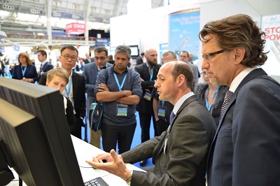
The CX Imaging Day provided a comprehensive programme of state-of-the-art imaging that is applied to the aorta, carotid, peripheral arteries and veins, including planning and using hybrid theatres, robotics, 3D fusion and guidance, intravascular ultrasound, perfusion angiography, cone beam computed tomography and carbon dioxide angiography.
This year’s Imaging Day was divided into two parts, giving attendees the opportunity for maximum learning and understanding. The morning session saw the presentation of the latest data in the Olympia Room Learning Centre, while in the afternoon, various imaging providers, including Philips, GE Healthcare, Siemens, Hansen Medical, Volcano and Ziehm Imaging, hosted “Ask the Expert” workshops at their stands in the Exhibition Hall. These workshops allow attendees to interact in small groups with the experts and to look at the imaging systems and tools, providing them with a unique view of the technology.
GE Healthcare
GE Healthcare took the opportunity to show the flow of its solutions from sizing to the 3D fusion from table side. There was also focus on dose optimisation management.
Alejandra Gonzalez, European marketing product manager for Interventional Systems at GE explained, “We provided an educational framework where we raised awareness of the key elements that physicians need to take care of in order to reduce the dose or optimise the dose strategy. We wanted to do it in an educational way and at the same time a more interactive way as well.”
Guiding the presentations and providing personal input were physician experts Stephan Haulon (Lille, France) and Adrien Hertault (Lille, France). Haulon explained what took place saying, “we showed the attendees a couple of cases that were sized on a workstation so they had the ability to actually manipulate the workstation and then we had them do a fusion registration to show them how straightforward a technology it is and how user friendly it is. We also had a big focus on dose exposure which is a real concern for vascular surgeons today because we are doing more and more radiation exposure procedures. We had a quiz which stirred a lot of questions and debates. So this was actually quite passionate and really interesting.”
Commenting on the workshop Haulon added, “There was a lot of interaction and those people would never stand up in a large room to ask a question, whereas here they were very enthusiastic about sharing their comments and asking us for more details so I think it was a very valuable workshop.”
Hansen Medical
Hansen Medical presented the Magellan robotic system which helps physicians navigate through the vasculature. Joe Guido, vice president marketing and business development explained that the system is designed to “make the procedure more efficient and more predictable for physicians and also allows physicians to stay out of the field of radiation. The robotic system will drive robotic catheters that you can shape and control without having to do exchanges for other catheters.”
Physician experts Barry Katzen (Miami, USA) and Celia Riga (London, UK) presented their experiences with intravascular robotics and the Magellan robotic system.
Guido commented on the workshop saying that many physicians are very curious about the technology and the practical applications and how it can benefit them and their practice. “An expert is much more accessible at the stand and it is a much more intimate environment. It encourages physicians to listen to the presentation and then speak to the experts and ask one-on-one questions,” he said.
Philips
Philips presented its technology and had physician experts Frank Vermassen (Ghent, Belgium) and Jim Reekers (Amsterdam, Netherlands) on hand to answer queries from physician-attendees. Philips demonstrators showcased the company’s technology including the VesselNavigator, a live image guidance solution; 2D Perfusion; Veradius Unity, a C-arm with flat detector; and gave advice on considerations when purchasing a hybrid room.
Commenting on the Veradius Unity, Philips demonstrator Marianne Kwakernaat said that the technology was developed, “to help get a more united team in the OR because there was a lot of frustration between doctors and operators who did not understand each other, so we made a lot of improvements to our systems to first, make it more easy to communicate, so we added some tools, and second, we made a new touchscreen which is very interactive and easy to use.”
She added that the workshop was a good vehicle to give the physicians the opportunity to see the products and figure out which one is most suitable to their hospital and their uses.
Siemens
Siemens showcased the complete workflow for EVAR procedures, starting with the preoperational CT scan. The company also gave attendees the opportunity to see the PURE software technology for the Artis zeego.
Dirk Sunderbrink, business manager, Therapy Systems at Siemens described the system saying, “It allows the vascular surgeon 2D and 3D registration with much less dose and in much less time than with previous systems. This PURE software guides the vascular surgeon through the procedure automatically to have fusion imaging available to guide them through the procedures.” He added, “The teaching opportunity is important to us because there are presentations, but it is much easier to really see it hands-on.”
Dittmar Böckler (Heidelberg, Germany) was on hand providing an expert position, giving attendees the opportunity to ask questions about the Siemens technology.
Volcano
Volcano showcased its intravascular ultrasound (IVUS) system, which provides detailed and accurate measurements of lumen and vessel size, plaque area and volume, and the location of key anatomical landmarks.
Physician expert Fabrizio Fanelli (Rome, Italy) was present to give his personal perspective of the technology and to answer attendees’ queries. At the stand, demonstrations were done showing real time sizing and procedure assessment with reduced exposure and contrast.
Volcano demonstrator Fiorella De Nicolais explained that the IVUS technology helps differentiate the four plaque types: fibrous, fibro-fatty, necrotic core and dense calcium.
She added that, “Not everyone knows IVUS, or the way it works or the benefits of using it, so the workshop is about getting more details and learning how the technology works.”
Ziehm Imaging
Ziehm Imaging featured its mobile hybrid room concept which consists of a mobile C-arm that has all the imaging capabilities of a fixed, installed hybrid room—high power imaging, a liquid cooling system, flat panel technology, fully motorised movements.
Visitors to the stand heard presentations from experts Wolfgang Keller (Nuremberg, Germany) and Peter Goverde (Antwerp, Belgium), who spoke about Ziehm’s mobile interventional suite and their clinical experiences with a mobile hybrid OR.
Summarising, Axel Kouril from Ziehm Imaging said, “As opposed to fixed installed hybrid ORs which are very expensive and have additional installations costs of enforcing the floor or the ceiling, and lead protection, this system has the size of a normal mobile C-arm and the imaging capabilities of a fixed installed system (up to 90%), but it is cheaper, it is mobile, and more flexible.”
A record-breaking week for the CX Venous Workshop
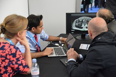
The second day of the CX Venous Workshop was held yesterday on the Upper Level of the Gallery, rounding off a busy week in which more than 950 people visited over the two days—a new record for the Workshop. Once again the educational stations, of which there were more than 30, were packed with delegates looking forward to one-on-one time with the distinguished Charing Cross Symposium faculty.
As was the case on Wednesday, the “Endovenous Ablation Village” was running, showcasing all available techniques to ablate truncal veins, along with practical training sessions teaching ultrasound-guided cannulation, catheter positioning and tumescent anaesthesia.
While the first day of the workshop focused largely on varicose veins and superficial venous issues, yesterday went deeper into the vein by examining acute deep vein thrombosis, intravascular ultrasound and deep venous stenting. Every station saw a significant amount of traffic, with delegates showing a keen interest in the intravascular ultrasound and deep vein thrombosis stations.
Ian Franklin, one of the course directors, said “Today we have had a big change of interest to deep venous—treatments of deep vein thrombosis, deep vein stenting, intravascular ultrasound, pelvic vein embolisation and so on—and it is packed. There is not a single empty training station, everyone has good interest and it is sustained. We deliberately have this flexible format so people can come and go—if they find they have a gap in the programme they can come and join us and make sure their time is used profitably.”
As the workshop drew to a close for another year, thoughts turned towards 2016. “Every year we change the event and it has never been the same as it was before, so next year will definitely be different,” said Franklin. “One thing I would consider is bringing in recorded live cases of all the new techniques we are showcasing to complement the hands-on demonstrations, using screens and some commentaries.”
With the workshop in its seventh year, there is always the opportunity to resurrect popular aspects of previous years’ Workshops. For example, Franklin suggested, “I would also like to bring back interactive discussion of complicated and difficult cases. That was always very popular, especially when you had a series of well-known experts all discussing how best to handle a case. Quite often we are showcasing different techniques and then once you have learnt that technique there comes the discussion of when to use it, so I think those case-based discussions complement the learning too.”
Day 2 offered stations covering: thermal (laser), thermal (radiofrequency ablation), non-thermal, practical training stations, valves, vascular ultrasound training simulator, venous malformations, pelvic venous imaging, pelvic vein embolisation, acute deep vein thrombosis, caval filters, follow-up protocols, air plethysmography, intravenous ultrasound and deep vein stenting.
Alma Lasers was exhibiting its VascuLife minimally invasive varicose vein laser treatment with robotic pullback. Having received CE mark and FDA approval recently, the VascuLife station had plenty of visitors looking to learn about the robotic pullback capability, which standardises pullback rate safely and allows a surgeon to focus on other tasks while pullback is completed. Shira Doron, marketing director at Alma Lasers, believes that the CX Venous Workshop gave companies and physicians a chance to interact and share information in an informal and purely scientific setting. She said, “Physicians view this as their professional platform as opposed to booths, which may feel like more of a marketing platform. They come here and it is all about the device and what it can do. You do not need to see movies or brochures—it is all about the technology.”
The new ArtVentive EOS device also was highlighted yesterday. Krzysztof Pyra (Lublin, Poland) spoke his experience with the EOS device treating ovarian veins for pelvic congestion syndrome. In addition, treatment of spermatic vein varicoceles were also presented. Pyra discussed his approach to diagnosing and treating pelvic vein incompetence. According to Pyra, of particular note was the immediacy of occlusion with the EOS device and shorter procedure times. Pyra uses a combination of EOS and sclerosant to occlude the pelvic veins.
Early results with a new thoracic branched device for the arch “are promising”
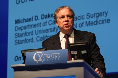
Michael Dake, Stanford, USA, presented the initial results with the GORE TAG Thoracic Branch Endoprosthesis (TBE), a new device for treatment of the aortic arch at CX yesterday.
Dake, Stanford, USA, presented the initial results with the GORE TAG Thoracic Branch Endoprosthesis (TBE), a new device for treatment of the aortic arch.
Dake told delegates that the GORE TAG TBE is a single branch device that consists of an aortic component, a side branch component and an optional aortic extender. The side branch component is delivered through a sheath and docks into the aortic component portal which allows for perfusion of a single arch branch vessel. The device is currently being studied in a zone 2 (left subclavian) feasibility study and a zone 0/1 (brachiocephalic and left common carotid) early feasibility study in the USA.
Currently, there are 20 patients (10 men and 10 women, mean age 75.4 years) successfully enrolled in zone 2 and one patient enrolled in zone 0/1. All 20 patients in zone 2 had successful access and deployment of the TBE device with the side branch component patent at the end of the procedure. Additionally, all side branch components remain patent. At one month, there have been no reported patient deaths or stroke. The left ankle brachial index (1.1) remains the same from pre-procedure to one month. There were no endoleaks that required reintervention.
The first two zone 0 patients were treated with the TBE device at the University of Pittsburgh Medical Center by Michel Makaroun, and at the Mayo Clinic by Gustavo Oderich. The protocol requires the patient to be treated in two phases with phase 1 revascularisation and phase 2 endovascular procedure. In phase 1, the patient underwent cervical debranching of the arch via a carotid to carotid bypass followed by a left carotid to left subclavian transposition. The proximal left common carotid and left subclavian artery was suture ligated.
Dake spoke about the patient treated about Makaroun and said the patient tolerated the procedure well without any neurological complications. The patient was assessed and deemed stable to proceed to phase 2 after 24 hours. In phase 2, the aortic component was advanced and deployed at the target location near the brachiocephalic artery. The sheath was advanced without difficulty into the portal of the aortic component into the brachiocephalic artery. The side branch component was advanced and deployed. Final arteriography showed excellent flow into the arch vessels, aneurysm exclusion, and complete apposition of the aortic component both proximally and distally.
Dake concluded: “These early results are promising with 100% technical success and side branch patency along with 0% death or stroke at one month.”