



CX 2026 Registration
CX Executive Board
CX 2026 Programme
CX 2026 – The 48th Charing Cross Symposium
The Charing Cross (CX) Symposium continues to set the standard in vascular and endovascular education, shaping the future of practice worldwide. CX 2026 marked the final chapter of our current three-year cycle, designed to spark debate on the biggest controversies in the field. Our distinguished faculty of global experts critically examined the evidence, exchanged perspectives, and worked towards building consensus on the issues that matter most.
Consensus was the defining theme for CX 2026. We welcomed an international community of specialists, educators, and innovators to tackle the pressing challenges in vascular and endovascular medicine—challenges that directly impact patient outcomes and clinical decision-making.
In 2024, CX found a new home at ExCeL London, unlocking fresh opportunities for immersive, hands-on learning. The expanded workshop programme has gone from strength to strength, offering delegates practical skills alongside groundbreaking evidence and innovation. In 2026, we returned once again to London’s premier conference and exhibition venue to deliver an agenda packed with cutting-edge science, expert debate, and future-shaping education.
Stay connected—sign up for the CX Newsletter and be the first to hear the latest updates.
CX Co-Chairs


Latest CX news
Fear of being sued may force healthcare providers into improving access to diabetic foot care services
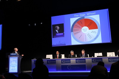
Although there is strong evidence that multidisciplinary foot care services can reduce the number of amputations associated with diabetic foot, access to such services remains patchy. Cliff Shearman (Southampton, UK) believes that healthcare providers are not doing enough to improve access. He says that it may take litigation from patients, who have lost a foot through poor care, to persuade healthcare providers to increase access, which he says would be “a great pity” because it should be “evidence and enthusiasm” that drives change.
Speaking at the CX ilegx Collaboration Day yesterday, Shearman explained that early diagnosis and prompt treatment can help to reduce the risk of amputation in patients with diabetic foot. He added that there is “strong evidence” to show that well-organised diabetic foot teams, working across primary and secondary care, can ensure that patients receive the care they need. Shearman noted that research indicates that such teams can the reduce rate of amputation by up to 80%. Furthermore, he reported that having a foot care team at his hospital (Southampton) has resulted in costs savings of £1,695,600 and a reduction in total beds of 5,662.
However, in the UK, only around 50% of people with diabetes receive the recommended nine health checks, including foot examination, and up to one in five hospitals do not have a diabetic foot protection team. Shearman commented: “I think healthcare professionals are now much more interested in the need to provide good care to reduce the amputation rates, but there is a lack of drive from government and healthcare providers to push this forward. At the moment, in the UK, we do not have core criteria for commissioning diabetic foot care services—all we can really do is to try to emulate those who have created good services,” he said.
According to Shearman, there are three steps to encouraging healthcare providers to take steps to increase access to foot care services: enthusiasm and evidence, naming and shaming, and litigation. He claimed that we are now the stage of “naming and shaming”—ie, showing how amputations rates can be “staggeringly reduced” with timely access and multidisciplinary team working. “If that does not work, it will be litigation that drives change. The number of ligation cases is rising enormously because people are recognising that they have not received good care and therefore, are taking action against their healthcare provider. It will be a great pity if litigation is what prompts change as it should be enthusiasm and evidence.”
Michael Edmonds (London, UK), who (like Shearman) is a course director of the CX ilegx Collaboration Day, agrees that poorly organised services—rather than a lack of effective treatment—are a cause of patients with diabetic foot having to undergo amputation. He said one of the key goals of ilegx, which was started in 2008, was to “encourage interdisciplinary collaboration that spans the primary and secondary services, and develop and implement best practice approach to save legs.”
Shearman told CX Daily News: “I hope delegates who attend the course will go away fired up with the knowledge that they can make a difference [by being enthusiastic about improving care and encouraging multidisciplinary working] and that they can help their centres save money as well as improve outcomes. Improving the care of patients with diabetic foot is not about hiring lots of new people or lots of new technology. It is about working as a team.”
Treatment options for diabetic foot
During the CX ilegx Collaboration Day, there were several talks about treatment options for patients with diabetic foot. Toby Richards (London, UK) spoke about a novel wound therapy called CelluTome (KCI). He said that this involved taking an epidermal graft off a patient’s leg, without any form of anaesthetic, by applying local heat to create microblisters that were then transferred onto a wound.
To assess the system, Richards and colleagues evaluated its use in a case series of 30 patients who were candidates for split skin grafts—most of whom had chronic wounds, with Richards noting: “so, we are not picking winners” (ie. patients with wounds that had a better chance of complete healing).
He stated that the “most important thing” was that the average time for donor site healing was 5.43±1.54 days, the mean pain score was 1.33±0.95, and the Vancouver Scar Scale was 0 for all cases at six weeks after therapy.
Richards reported that 17 patients had complete healing (12 in under six week and four in under eight weeks) and that seven grafts failed due to infection, adding “one of the problems we had is that the graft looks like a biofilm at two weeks and a nurse who was not familiar would come along and think it needed cleaning, so there goes your graft. While you get used to this look, you need to leave the dressing on as long as possible, and then you will start to get good results.” He concluded that it was a novel technology that was “feasible” and it that “certainly does work”, saying: “as with all new technologies, it should be tested in a proper trial before we introduce it into normal practice. Therefore, we will be evaluating it in a randomised controlled trial.”
Jennifer Tremlett (London, UK) also spoke about wound care techniques, including platelet rich plasma, larvae therapy, and negative wound pressure. Like Shearman and Edmonds, Tremlett advocated “multidisciplinary working” because it “provides better outcomes”. Furthermore, Dean Huang (London, UK), who spoke about advances in distal endovascular surgery, said that the need for collaborative working and rapid access to diagnosis and intervention were “probably two of the most important points” in his talk.
“We can solve the limitations of carotid stenting with a micro-mesh stent and its sustained anti-embolic action”
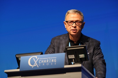
Micro-mesh stents and sustained anti-embolic action to reduce cerebral embolisation may contribute to solving the remaining limitations of carotid stenting, according to Alberto Cremonesi (Cotignola, Italy), who spoke at CX 2015 (28 April–1 May, London, UK).
Cremonesi examined the potential for micro-mesh stents to improve existing carotid artery stenting outcomes.
He told delegates that the correct technique for performing a carotid artery stenting was to conduct a pre-procedural evaluation for common carotid engagement, to select the correct stent and to select and manage an embolic protection device.
Cremonesi said that it is already known that “not all plaques are the same”, and that “carotid artery stenting and carotid endarterectomy are —and will remain—emboli-generating procedures”. He told the audience that it is possible to give sustained anti-embolic over time using specific stents.
When a poorly-selected stent is used for carotid artery stenting, intra-strut prolapse can occur resulting in post-procedural embolic events. The difficulty then, commented Cremonesi, is that “lesions in the carotid arteries are often anatomically and morphologically very challenging. For this reason, plaque coverage can play an important role.”
The use of stenting strategies such as open cell, closed cell and hybrid geometry can be considered “time honoured”. Now, vascular specialists have access micro-mesh double layer carotid stents (Terumo, InspireMD and Gore), said Cremonesi, whose personal experience is with the Roadsaver carotid stent from Terumo.
Cremonesi admitted that “In terms of data, we do not have very much at the moment” for the use of micro-mesh. Some data do exist from the CARENET all-comer trial, in which 30 patients saw “very good” outcomes without any neurological events up to five months follow-up. A further multicentre trial with 100 patients, CLEAR-ROAD, is also beginning, though Cremonesi said that he did “not want to conclude anything” about it yet.
“We are probably changing the paradigms in carotid artery stenting, because with these micro-mesh stents and their sustained anti-embolic action, we can solve the remaining limitation of carotid stenting,” ended Cremonesi.
Discussing his presentation with the audience, Cremonesi mentioned that “two-thirds of the embolic complications are in the post-procedural phase.” When session chairman Roger Greenhalgh asked about the cause for this later onset of complications, Cremonesi told him that it was due to the stent interacting with and cutting the plaque in the carotid artery, which can occur during the phase and even up to 48 hours later. Cremonesi made it clear that this problem is one of the carotid artery, rather than one associated with the aortic arch.
Despite endovascular revolution, 65% say heyday of open repair in aortic arch continues
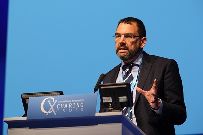
There have been vast technological advances that have enabled the final frontier of the aorta, the diseased ascending aorta and arch, to be treated by completely endovascular means. Simultaneously, there have also been advances in open repair. Also, with perioperative stroke remaining a principal risk with TEVAR, the high incidence of cerebral embolisation with the procedure is a problem that needs wider recognition, delegates heard yesterday.
In a panel discussion during the session on interventions for ascending aorta and aortic arch, Roger Greenhalgh, chairman of the CX Organising Board, pressed the panel to comment on patient mortality after open repair and endovascular repair.
The mortality rate for open repair is around 3%, even when there is involvement of the aortic valve, said Stephen Large, Cambridge, UK. With endovascular repair, it approaches 10%, said Dittmar Böckler, Heidelberg, Germany. Subsequently, 65% of the CX 2015 audience voted against the motion that the heyday of open aortic surgery is over.
Richard Gibbs, London, UK, told CX delegates yesterday that there was a high rate of embolisation during arch and descending thoracic intervention that could be observed as silent cerebral infarction on new imaging techniques. The results from the study he presented showed that there is a 70% silent cerebral infarction on MRI and that there is postoperative neurocognitive decline in patients with silent cerebral infarctions.
Gibbs made the point that stroke, which was caused by cerebral embolisation, was a relatively crude surrogate, but a clinically relevant measure of microembolisation. The stroke rate for TEVAR ranges between 3% and 6%, said Gibbs, with embolisation being caused by the passage of stiff wires, soft wires, devices and manipulation within the diseased aorta. The risk factors are the atheromatous burden within the aorta and using a proximal landing zone. Hypotension is also very important, he said.
“A much subtler way of looking at microembolisation is with diffusion-weighted MR that detects acute ischaemia that is due to cerebral oedema. These lesions appear within 24 hours of the insult and last up to 14 days, and show as hyperintense bright areas that are easy to recognise,” Gibbs noted.
Gibbs then showed diffusion-weighted cerebral MR images from a patient who had silent cerebral infarctions but did not develop signs or symptoms of clinical stroke following TEVAR. “Therefore what we see here is silent stroke, or silent cerebral infarction, which is imaging evidence of cerebral infarction, without a history of acute neurological dysfunction attributable to the lesion,” Gibbs said.
He qualified that he believed the term silent cerebral infarction is a misnomer because there is an increasing body of evidence that suggests that silent cerebral infarction is associated with depression, dementia, Alzheimer’s, future increased risk of stroke and mortality.
Published literature shows a significant rate of cerebral infarction with various different interventions involving the arch and unsurprisingly transcatheter aortic valve implantation (TAVI), which is the biggest device and causes the most cerebral infarction, he explained.
“When it comes to TEVAR, there is very little literature, and one paper suggests that there is a 63% risk of cerebral infarction with the procedure,” said Gibbs referring to the data from 19 patients from Kahlert et al published in the Annals of Thoracic Surgery in 2014 that found diffusion-weighted MR evidence of cerebral infarction after TEVAR in 12 of 19 (63%) undergoing the procedure for a variety of indications. There was no overt clinical stroke seen in these patients.
Gibbs then presented the pilot work done by his team on the rates of silent cerebral infarctions during TEVAR, the presence of silent cerebral infarction on MR and whether there are neurocognitive changes afterwords.
“We included 44 patients undergoing TEVAR and looked at the burden of atheroma within the arch of the descending aorta based on the American Heart Association grading method. The patients had bidirectional transcranial Doppler, which is a direct measure of cerebal microembolisation. “A subset of these patients had pre-and postoperative MR looking for more evidence of infarction (23 patients). Another subset had neurocognitive assessment before the intervention, after the intervention in hospital and eight weeks later to see if any changes persist,” said Gibbs.
Forty one TEVAR procedures involving the arch and descending aorta were performed for a variety of pathologies. Of these 21 were standard and 20 were complex involving the use of branches, scallops or adjunctive surgical procedures. “We had fairly proximal landing zones,” he noted.
“Looking at the specific procedural steps, we see clearly that stent graft deployment carries the most significant rate of embolisation,” said Gibbs. He then showed a transcranial Doppler during stent graft deployment where a burst of cerebral embolisation was visible in both hemispheres. “The highest activity of cerebral embolisation was when the device was deployed. Increased embolisation was associated with the left hemisphere more than the right; stent manipulation more than wire and catheter passage; a higher grade of atheroma compared to a lower grade (so a higher burden of disease); chronic rather than acute disease. Patients who had a stroke had the highest rate of embolisation. A proximal landing zone rather than distal one had a higher rate of cerebral embolisation. In the group where we looked for evidence of silent cerebral infarction, 70% (16/23) of our patients had this, mostly in the left hemisphere and mostly in the territory served by the middle cerebral artery. Ten per cent of these patients had a clinical stroke,” said Gibbs.
When the researchers selected out the data for older patients from the group, they found that executive function diminished and stayed down at eight weeks, memory diminished and manual dexterity got worse.
“There is a significantly high rate of cerebral embolisation during TEVAR and the more proximal you go, the worse it is. There is a definitive and radiologically proven damage to the brain and these patients do pay a price for this. We have to be thinking about how we can intervene [either pharmacologically or by using devices],” concluded Gibbs.
Scarcity of literature on proximal landing zone and TEVAR outcomes
Dittmar Böckler stated that while the relationship between the proximal landing zone and outcome was well-documented in the EVAR literature, there were very few publications focused on how the proximal landing zone influences patient outcomes with TEVAR in the arch, and noted the low level of evidence.
“There are no randomised controlled trial data on open versus endovascular approaches and there are no meta-analyses. The data come from limited European registry data from the Relay registry, Traviata registry and European CTAG registry,” he noted.
Böckler drew attention to the fact that there was no consensus in the literature and guidelines on the appropriate proximal neck length and that this variation was reflected in the instructions for use from manufacturers, as the target landing zone is depending on stent graft diameter.
“The appropriate proximal landing zone for TEVAR is not defined. Arch type and atheroma seem to influence stroke risk during TEVAR in the arch. “There is strong need for new refined conformable devices in the arch including branched stent graft technology,” Böckler said.
Value of medication
Frank Lederle, Minneapolis, USA, speaking on the value of medication such as statins before aortic arch catheterisation, stated that there was very little trustworthy randomised controlled trial data to go by. He reviewed evidence that bears on whether perioperative statins or aspirin benefit patients having a thoracic aortic procedure. “Analyses showing benefit of perioperative use rely on weak and doubtful studies. When it comes to aspirin, there are no data on perioperative benefit, but there is an increased major and minor bleeding,” he said.
Lederle drew attention to the fact that many patients with aortic aneurysm have arteriosclerotic cardiovascular disease and should be on statins and aspirin long-term. “There is no good evidence that statins or aspirin improve long-term outcomes for patients with aortic arch patients without arteriosclerotic cardiovascular disease, so some of these patients will not be on these medications. Questions remain on when whether or not patients are on them long-term and whether they should be used perioperatively,” he said.
Open surgery
Stephen Large, Cambridge, UK, a cardiac surgeon, outlined the case for open surgery in interventions for ascending and arch of the aorta, the current gold standard approach.
Large noted that not operating resulted in a “dreadful attrition”. “We know that there is an increase in attrition correlated with the increase in aneurysm size, the hingepoint being around 5.5cm in the ascending and arch, which is associated with an acceleration in terms of stroke, dissection rupture and death,” he said.
“What we very often do in the ascending aorta is deal with an associated post-aortic valvular lesion either by an interposition graft, that is something placed above the coronary artery ostia, really at the level of the sinotubular junction, and right up to the origin of the innominate artery—a true ascending aortic replacement. We can, if we are in trouble (of course with involvement of pathology within the aortic root), replace the aortic root and that always requires reimplantation of the coronary ostia, which brings a raft of problems of threatening ischaemia with it. Up until relatively recently, this involved automatic replacement of the aortic valve. There is now a keen interest in considering valve preservation procedures, something that I have fought against for many years, because the aortic valve is embryologically of the same origin as the ascending aorta. However, counterintuitively, it appears that the aortic valve fares very well. So what to do with the arch? We can replace it with a tube graft and address each of the usual three arch vessels. Or we can translocate the whole of the aortic arch vessels permitting the use of either replacement or stenting. As cardiac surgeons, we find ourselves replacing the ascending aorta in an emergency as a life-saving procedure for dissection. We, of course, will look at ascending aortic aneurysms for elective surgery for prognostic issues and this is often in conjunction with other procedures in the chest such as aortic valve replacement,” he said.
Endovascular procedures a valid alternative in selected patients
Piergiorgio Cao, Rome, Italy, speaking on branched stent grafts for the treatment of complex arch lesions, said that any repair of aortic arch remains demanding and exposes patients to mortality and stroke risks that are “not negligible”. Open repair is the gold standard, and hybrid and endovascular repair are valid alternatives, mostly in patients who are at high risk for surgery, he noted.
Cao defined the morphological feasibility to receive endovascular treatment as the presence of a proximal landing ≥2cm in length and ≤4.2cm in diameter. The challenges for TEVAR in the arch include conformability of the stent graft, endoleak and retrograde dissection occurence, he said.
He then alluded to a recent publication in the Journal of Vascular Surgery from Paola De Rango et al that analysed total aortic arch reconstruction in a contemporary comparison of current open and endovascular repair.
De Rango et al entered endovascular and open arch procedures performed from 2007 to 2013 into a prospective database and then retrospectively analysed the data. Endovascular repair (proximal landing zones 0-1), with or without a hybrid adjunct, was selected for patients who were sicker but who had a fit anatomy. Operations involving coverage of left subclavian artery only (zone 2 proximal landing) and open hemiarch replacement were excluded.
As reported in the journal, the authors concluded that despite the older age and a higher comorbidity profile in patients with challenging aortic arch disease who were suitable and selected for endovascular arch repair, no significant differences were detected in perioperative and four-year outcomes compared with the younger patients undergoing open arch total repair.
Yesterday, Cao concluded by saying: “The endovascular approach is a valid alternative to open surgery for all patients, when morphologically feasible. A safer proximal landing zone with longer coverage of the ascending aorta may be the key for long-term durability and to prevent retrograde dissection. Branched stent grafts might be useful in avoiding arch manipulations and decreasing the risk of major adverse events,” he said.
Greenhalgh then commented that the audience might like to have some idea of the expected mortality associated with both the open and endovascular methods in the case of a 70-year-old patient with an ascending dilating disease process in which the valve becomes incompetent. Mortality is 3% with open repair and 10% with endovascular repair, the audience learned.
He further commented on the resurgence of the classification of aneurysmal disease: “The starting point seems to be aneurysmal disease. We are seeing comments on syphilitic, fusiform and saccular aneurysms and this is beginning to look like an old surgical textbook, he commented.
In a debate, Hans-Henning Eckstein, Munich, Germany, argued against the motion “The heyday of open aortic surgery is over” to garner majority support that open repair still had a valid place in the treatment of the ascending aorta and aortic arch. He persuaded 65% of the delegates to vote against the motion. Frank Veith, New York, USA, spoke for the motion.