

CX NEWS
CX Imaging Day: High-quality imaging is the key to optimal results
Stéphan Haulon, Richard McWilliams and Alan Lumsden, Faculty members of the CX Imaging Day, new style Parallel Session of the Charing Cross Symposium 2015), outline the relevance of therapeutic imaging for vascular and endovascular procedures, the use of robotics and radiation exposure concerns. These topics will be discussed at the CX Imaging Day on Thursday 30 April.
This year, the CX Imaging Day will provide a comprehensive programme on state-of-the-art imaging applied for the intervention of the aorta, carotid arteries, peripheral arteries and veins, involving planning and using hybrid theatres, robotics, 3D fusion and guidance, intravascular ultrasound (IVUS), perfusion angiography, cone beam computed tomography (CT) and carbon dioxide angiography. In the morning, attendees will hear the latest data on high-quality imaging and be able to discuss the different applications. During the afternoon, the “Ask the expert” workshop is organised in the Exhibition Hall with visits to the industry stands, focusing on sizing, 2D/3D fusion imaging and how to achieve optimal dose levels.
Stéphan Haulon (Aortic Centre, Hôpital Cardiologique, CHRU Lille, France), says delegates will get insights from large cohort published studies to identify the various strengths and limitations of high-end imaging. He adds: “We will report the latest progress, provide an update on radiation baseline for various procedures, and see how these new imaging techniques can be extended to peripheral limbs.”
Richard McWilliams (Radiology Department, Royal Liverpool University Hospital, Liverpool, UK) notes that the immediate and continuing success of endovascular interventions relies heavily on medical imaging. He comments: “The processing power of modern imaging workstations allows the physician to rapidly assimilate the relevant anatomy before intervention. Procedural success is facilitated by the quality of modern hybrid suites and the associated software that accompanies these allowing 3D imaging, 3D fusion and 3D guidance. Surveillance after endovascular treatment is now of higher quality because of improvements in CT scanners and ultrasound techniques including contrast-enhanced ultrasound. Direct comparison of one CT after aneurysm repair with a prior CT is now a joy when these are registered and locked together on dual monitors and reviewed in cine mode.”
Haulon states that the rise of catheter-based procedures and minimally invasive surgery has required concomitant advances in intraoperative imaging applications, and that hybrid rooms have made this possible by combining the mobility and sterility of an optimal open surgical environment with the latest advanced imaging capabilities of a fixed system such as image fusion or cone beam CT. “Experience has shown that the routine use of advanced imaging techniques, such as fusion, during endovascular aneurysm repair has significantly reduced the radiation exposure to patients and operators, as well as the contrast volume load, without jeopardising the overall procedure workflow,” he says.
The role of intraoperative imaging will be also highlighted in the Main Programme – Abdominal Aortic Controversies session (Wednesday 29 April) with a presentation by Dittmar Boeckler on intraoperative CT in ruptured abdominal aortic aneurysm treatment with EVAR.
The portability of imaging (eg. ultrasound) and the more widespread availability of CT and MRI scans have transformed diagnosis and now the treatment of vascular disease, according to Alan Lumsden (Houston Methodist Hospital, Houston, USA). “We are in an era where portability makes imaging technology available in the operating room—image-guided surgery and endovascular surgery. This can be either by direct imaging or increasingly the use of image fusion methodologies.
At the CX Imaging Day, Lumsden will show how the use of robotics can be beneficial for different procedures—carotid, complex cases and even retrieval of inferior vena cava filters. “Navigation through the vascular system depends on the use of wires, catheters and a series of wall interactions with these devices. Such techniques have not fundamentally changed in decades. Catheter robotics promises a level of catheter and wire control that simply cannot be achieved using traditional techniques,” he says. Lumsden explains that his group’s interest in robotics was generated by watching an electrophysiologist take the robotic catheter through the inter-atrial septum, in a beating heart, while applying measured pressure and radiofrequency energy at multiple points, under precise control to the wall of the left atrium. He adds, “It was hard not to be impressed with these capabilities, hence our interest in the peripheral application of robotics. We believe that controlled navigation, stability and pushability of the Magellan catheter (Hansen Medical) will permit us to perform procedures which cannot be completed using manual techniques. Further it will allow us to steer devices which lack sterility (snares, thrombectomy devices, atherectomy catheters), thereby improving their capabilities.”
Haulon will present a new integrated workflow for sizing, 2D/3D fusion and assessment of endovascular aneurysm repair (EVAR). He explains: “Time is crucial, improving vascular surgeon sizing and fusion workflow is key, so why not preparing both steps at the same time? Completion contrast-enhanced cone beam CT has also demonstrated its capacity to assess technical success, reducing the requirement for a postoperative CT angiography and allowing accurate detection of complications requiring additional treatment right on the spot. Also, there is a need to consider radiation dose at a higher level than that of the procedure itself… we need to take into consideration the patient’s dose throughout the entire treatment process, from diagnosis, to treatment and including follow-up. Having the right dose management tools in place allows me to achieve this.” Giuseppe Pannuccio (Muenster, Germany) will be also presenting on the role of 2D/3D fusion imaging in complex aortic procedures.
3D imaging will be further explored by Tara Mastracci (London,UK) who will discuss the role of 3D guidance in fenestrated endovascular aneurysm repair (FEVAR) and by Frank Vermassen (Ghent, Belgium) who will show how 3D imaging can be used in carotid procedures. Jan Brunkwall (Cologne, Germany) will focus on 3D imaging in complex aortic procedures.
Radiation exposure concerns
Radiation protection remains a major concern and will be one the topics discussed at the CX Imaging Day, and new technology is helping to change behaviour and lower doses, says McWilliams, and adds: “Live dose monitoring provides instant feedback to operators and assistants so that they are aware how their actions increase or decrease dose. Newer equipment is increasingly being designed with dose-saving in mind. Intravascular ultrasound during venous procedures is an important part of this.”
He maintains that “we must also remember the simple things that reduce dose which I see forgotten very commonly. These include increasing the distance from the X-ray source, using face screens, using pump injections rather than hand-injections to allow the operator to increase his/her distance, reducing the pulse rate and frame rate and knowing the relevant projections from the preoperative dataset rather than by trial and error during the procedure. An unfortunate reality is that although modern equipment is capable of exquisite fluoroscopic images, the operator needs to ensure that the images are good enough but not needlessly better than this, to avoid excessive dose to the patient and staff. If the image quality is too good then this is bad”.
Radiation exposure will be also highlighted in the Main Programme – Abdominal Aortic Controversies session with a mini-symposium dedicated to explore radiation reduction strategies for the operator and the patient, including a presentation on dose optimised imaging protocols for complex endovascular procedures by Eric Verhoeven (Nurnberg, Germany). In the Peripheral Arterial Controversies session (Tuesday 28 April) of the Main Programme, Martin Malina (Malmo, Sweden) will be speaking about contrast reduction angiography for peripheral arterial disease.
In the afternoon, delegates will visit the Exhibition Hall (GE Healthcare, Hansen Medical, Philips and Siemens booths) for technology demonstrations.
The New CX Imaging Day will take place at the Charing Cross Symposium on Thursday 30 April – Olympia Room Learning Centre in the morning and Exhibition Hall in the afternoon, Olympia Grand, London, UK.
Click here to see the CX Main Programme Sessions
Click here to see the CX Imaging Day
Click here to register
Vascular malformations and limb ischaemia: Main topics of the CX Paediatric Vascular Issues session 2015
In 2015, the CX Paediatric Vascular Issues session will host presentations on modern management of childhood vascular disorders with a special focus on vascular malformations and limb ischaemia as well as other pathologies (aortic and arterial). Course directors, Malcolm Simms (Birmingham, UK) and George Hamilton (London, UK), outline the hottest controversies in the field.
Simms says that there are at least three areas of controversy when treating children with vascular issues, “the appropriate management of iatrogenic acute limb ischaemia, the optimal management of vascular malformations and the tailoring of vascular intervention in children to address issues of growth”.
Adding to the current controversies in the field, Hamilton comments: “There is a very low level of training or at the very least understanding of available treatments and protocols in current vascular training programmes throughout Europe. As a result poor treatment and outcomes are common.” He adds, “Paediatric vascular treatment should be delivered in specialist children’s centres.” However, there is still a need to develop such centres to deliver optimal care, he notes.
With regards to improvements in the field, Simms states that the most relevant advance has been the development of smaller calibre angiography catheters and devices. “This has extended the scope of endovascular diagnosis and intervention to smaller children.” Hamilton comments that another relevant advancement is the “growing expertise and confidence in the use of thrombolysis in acute ischaemia with the huge potential to avoid amputation and save limbs.” The understanding of vascular malformations has also been improved with better nomenclature and classification, Simms adds.
The course directors note that acute limb ischaemia treatment is the area that urges most attention in paediatric vascular treatment. Even though there are some advances in the field, there is still a special need to further develop (particularly for children younger than five) small diameter endovascular catheters, wires and absorbable small diameter stents, which seem to offer the best potential for treatment because of growth issues, Simms and Hamilton comment. Simms adds that the management of large diffuse vascular malformations remains “palliative at best”.
Delegates who attend the CX Paediatric Vascular Issues session will learn about the latest treatment methods in the field and will also have the chance to discuss specific examples with case-based presentations.
The CX Paediatric Vascular Issues session will take place at the Charing Cross Symposium on Thursday 30 April (morning) – London Room Learning Centre, Olympia Grand, London, UK.
Click here to see the CX Main Programme Sessions
Click here to see the CX Paediatric Vascular Issues session
Click here to register
Call for case submissions to the CX Paediatric Vascular Issues session
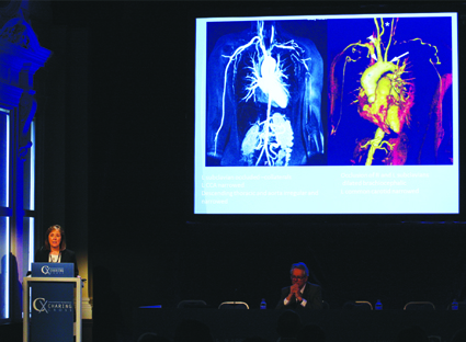
The Charing Cross Symposium is looking for paediatric vascular management dilemmas to encourage audience discussion at the CX Paediatric Vascular Issues session.
Clinicians are invited to submit paediatric cases concerning acute or chronic limb ischaemia, trauma, mid-aortic syndrome, cancer resection and vascular malformations.
Format: Cases should be submitted in Powerpoint (maximum three slides or the equivalent to a three-minute presentation). The material should be limited to the case presentation for discussion by the delegates and expert panel – no ‘lectures’ or literature review of the associated condition should be included.
Deadline for submission: 17 April 2015
Please send your cases to: Katherine Pole (Katherine@bibamedical.com)
Get the latest update on vascular malformations management
Delegates attending the CX Vascular Malformations Management session on Thursday 30 April, in the afternoon, will get a general overview of congenital vascular malformations and will also learn about the latest advancements in the field.
This year, the course will include keynote lectures covering the topics of venous, lymphatic, combined and arteriovenous malformations each followed by case presentations illustrating representative clinical situations. The role of imaging of vascular malformations and treatment options including embolisation, sclerotherapy and laser therapy will be addressed.
The session, led by Iris Baumgartner, Bern, Switzerland, will be interactive allowing the audience to get feedback from a panel, which includes the experts: Raul Matassi, Matthias Widmer, Joe Brookes, Andreas Saleh and Martin Köcher.
The CX Vascular Malformations Management session will take place at the Charing Cross Symposium on Thursday 30 April (afternoon) – London Room Learning Centre, Olympia Grand, London, UK.
Click here to see the CX Main Programme Sessions
Click here to see the CX Vascular Malformations Management session
Click here to register
Four days of Venous content to be run for the first time at Charing Cross
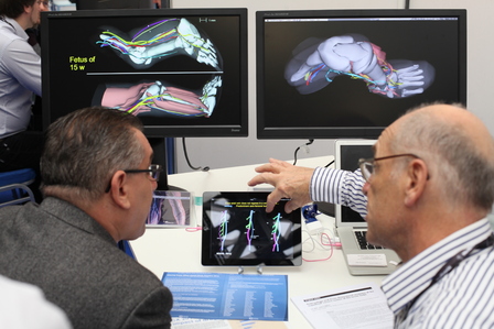
In 2015, the Charing Cross Symposium will dedicate, for the first time, four days of discussion in the venous field addressing both the superficial and deep venous systems. The Symposium is also launching a new CX Venous Workshop, formerly named as CX Office-Based Vein Practice Course, which includes hands-on training on the latest techniques for varicose veins treatment and, for the first time, practical training on deep venous treatment.
On the first day (Tuesday, 28 April) upcoming leading vascular and endovascular experts will present cutting-edge venous investigation at a CX Abstracts‒Venous session. Delegates will also have the chance to visit the CX Venous Workshop, which will run on the following days (Wednesday, 29 April and Thursday, 30 April). On the fourth day, the CX Main Programme will be dedicated to Venous Controversies.
Superficial and deep venous treatment at the core of the Venous Controversies
This year’s Venous Controversies in varicose veins treatment are no longer between surgery and office-based techniques, according to Roger Greenhalgh, (Imperial College, London, UK), chairman of the CX Programme Organising Board. “It seems as if office-based vein techniques have swept the board. Now the controversy seems to be about which method to select in which situation,” he says.
Delegates will learn the latest evidence on varicose veins treatment options with thermal and non-thermal techniques. “We will try to present a balance between data and information that relate to office-based tested techniques that are already established and are probably state-of-the-art alongside techniques that are emerging, and that may not have enough data but nonetheless might represent a completely shift in practice. We are happy for people to find out about these new techniques at Charing Cross first,” says Ian Franklin (London, UK), member of the CX Programme Organising Board.
Commenting on non-thermal techniques, Mark Whiteley (University of Surrey, London, UK), also a member of the CX Programme Organising Board, says: “The newer non-thermal techniques are now joining the endovenous thermal ablative devices and starting to show impressive results. The controversy here will be whether these newer non-thermal methods are now able to replace endothermal ablation in some or all cases.” (See an interview with Whiteley on varicose veins treatment).
Sclerotherapy and laser therapy of cosmetic skin problems will also be discussed.
Deep venous treatment is another major topic of controversy at this year’s CX Symposium. Franklin comments: “Deep venous thrombosis treatment is controversial because it is extremely badly managed in many countries and some people get almost no treatment at all. Nowadays, there are new technologies that help physicians treat deep venous thrombosis effectively. There is a lot to achieve in terms of training and education on how to diagnose deep venous thrombosis promptly with the right method of treatment to guarantee durable results.”
Stephen Black (Guy’s and St Thomas NHS Foundation Trust, London, UK), member of the CX Programme Organising Board, considers that there is a real need to develop the evidence base for treatment options for deep venous thrombosis. At the session on Deep Venous Controversies, delegates will learn about the latest evidence on the current techniques for deep venous thrombosis treatment including new stents and catheter-directed thrombolysis and thrombectomy techniques. (See an interview with Black on deep venous disease treatment).
Imaging for superficial and deep venous treatment is playing a vital role in the diagnosis of these anomalies. Greenhalgh comments: “It appears that imaging is absolutely at the core of intervention in the superficial and deep venous systems. Unfortunately, venous imaging is less than helpful when the patient lies flat and how to image the deep venous system with the patient standing will be discussed.”
The use of current imaging technologies such as duplex, magnetic resonance imaging (MRI) and intravascular ultrasound (IVUS) will be discussed at a session dedicated to this field.
New CX Venous Workshop
The former CX Office-Based Vein Practice Course has changed its name to the CX Venous Workshop because it now encompasses all aspects of venous disease including deep vein thrombosis, intravascular ultrasound and deep vein stenting. The course will continue providing delegates with hands-on training sessions for varicose veins treatment. In 2014, it had a record attendance of over 700 delegates during two days.
The CX Venous Workshop is set out in a very flexible format and will run from 10am until 3pm over two days. There will be different training stations (around 30) led by a physician expert who will be demonstrating a wide range of techniques and procedures in different superficial and deep venous treatment techniques. There is not a rigid timetable and participants can choose their timing at their convenience. The course facilitates one-to-one interaction or discussions in small groups.
Key areas covered include:
- Practical training
- Truncal ablation
- Diagnostic venous ultrasound
- Anatomy
- Venous haemodynamics
- Sclerotherapy
- Thread veins
- Dermal lasers
- Pelvic venous imaging
- Pelvic congestion
- Pelvic veins and haemorrhoids
- IVUS
- Deep vein stenting
- Pelvic vein embolisation
- Lymphoedema
- Endovenous glue
- Mechanochemoablation
- Steam
- Wound care
- Varicose veins
- Perforators
- Tributaries
- Surgery
- Caval filters
- Intermittent pneumatic compression
- Stockings and bandaging
- Neuromuscular electrical stimulation
- Thrombophilia and DVT Venous malformations
The CX Venous Controversies Day will take place at the Charing Cross Symposium on Friday 1 May – Main Auditorium, Olympia Grand, London, UK.
Delegates will have the opportunity to get hands-on training on the latest techniques for superficial and deep venous system treatment at the CX Venous Workshop on Wednesday 29 April and Thursday 30 April – Gallery Upper Level, Olympia Grand, London, UK
Click here to see the CX Main Programme Sessions
Click here to see the CX Parallel Sessions
Click here to see the CX Venous Workshop
Click here to register
Insights into the current practice for varicose veins treatment
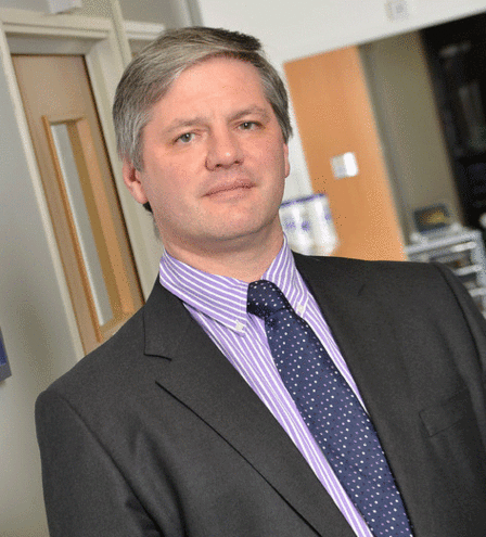
Venous surgeons and phlebologists are spoiled for choice when it comes to office-based technology options for varicose veins. This brings questions such as what technologies are best suited for what patients, what factors should be considered to obtain best results and what is the role of open surgery in the field. Mark Whiteley (University of Surrey, London, UK), member of the CX Programme Organising Board, gives his views on the subject. The latest evidence in this area will be discussed in the Venous Controversies sessions of this year’s CX Main Programme.
Physicians now have a great selection of office-based technologies for the treatment of varicose veins; what method is best for what case?
This has been the major area of my research since starting radiofrequency ablation in March 1999 and it is fair to say that there are many different opinions on this. Most importantly, before this question can even be considered, we need to decide on what constitutes “the best”. In my view, technical perfection is the correct outcome, as this will lead to long-term success and therefore should lead to long-term patient improvement.
However, advocates of techniques with inferior technical results that might require less intervention or fewer injections, point to patient satisfaction as the correct outcome measure to use to assess “the best” treatment method. I believe this is not a good outcome measure to use scientifically. Patient satisfaction can be manipulated by factors other than the treatment itself, particularly by misinformation given to patients in some studies by advocates of one method as to the theoretical risks of other methods that might be used.
In addition, satisfaction has to be measured at one or several points in time—often in the relatively short term. Technical imperfections which are likely to progress to recurrence and reduced patient satisfaction in the long term might be present in the short term. But measuring patient satisfaction alone may miss these out due to the time it takes for the patients to experience the clinical outcomes of the treatment failures.
What factors should be considered to obtain best results in varicose veins treatment?
As with all surgical techniques, there are several factors involved in obtaining the best results. Not only does the technology and device design have to be adequate to the job, but the patient selection needs to provide appropriate cases for the chosen technique and doctors and their teams need to use adequate imaging and procedural technique to get the optimal results.
In addition, patient expectation has to be added, such as whether they are looking for relief of symptoms only or cosmetic perfection and also an acceptable cost for the service.
What office-based technologies are currently showing best results?
Generally, in most hands, endovenous thermoablation shows excellent results for truncal and perforator veins ablation, provided the correct technique is used and an appropriate device for the vein to be treated. Glue and mechanochemical endovenous ablation (MOCA) are starting to show promising results in truncal venous reflux in selected patients whereas foam sclerotherapy is technical disappointing in truncal veins. However, advocates will point to patient satisfaction and re-treatments to argue for its use.
Currently, coil embolisation of refluxing pelvic veins with foam sclerotherapy seems to be the optimal treatment for pelvic venous reflux. Foam sclerotherapy alone or ligation of veins at the pelvic exit points are used by some but need to be proven as effective and acceptable treatments.
For incompetent tributaries and varices, ambulatory phlebectomy is probably optimal for larger veins and foam sclerotherapy for smaller veins— although the new technique of combining both as “foam phlebotomies” is gaining popularity in the USA.
When it comes to neovascular tissue, strip tract revascularisation, feeding veins under patches of telangiectasia and primary avalvular varicose anomalies (PAVA) then foam sclerotherapy is the only effective option.
Finally, for telangiectasia themselves, microsclerotherapy is still the best option as it treats both the visible veins as well as the communicating veins hidden below the surface.
With the outcome of many office-based technologies and their comparable efficacy results with surgery, is there still room for surgery?
No.
Unless one calls ambulatory phlebectomies (surgery), then there is no need to ever perform open surgery for varicose veins anymore. Personally, I have not had to use open surgery for any varicose veins case for over a decade.
At the debate “Duplex scanning is mandatory before treatment of asymptomatic cosmetic thread veins” you will present “For the motion”. Could you tell us the reasons as to why you are supporting this practice?
Since 2001, increasing numbers of research papers have shown that cosmetic thread veins frequently occur in association with underlying venous reflux. In many, if not the majority of cases, this venous reflux leads directly into the thread veins. Furthermore, every case of the complication called telangiectatic matting that I have ever seen after thread vein treatment, has an underlying untreated vein refluxing into the affected patch.
Since instituting a policy of duplex scanning in 2002 in which every patient presenting with cosmetic thread veins of the legs are treated for any underlying venous reflux before proceeding to microsclerotherapy of the thread veins themselves, we have never had a case of telangiectatic matting. Those cases presenting to us with this complication from elsewhere invariably have an underlying vein refluxing into the lesion.
With the explosion of cosmetic clinics starting to offer thread vein treatments, often without duplex ultrasound or even hand held Doppler, it is essential for us as phlebologists and venous surgeons to state that we feel that treatment of thread veins without a duplex ultrasound scan first is a sub-optimal treatment.
The CX Venous Controversies Day will take place at the Charing Cross Symposium on Friday 1 May – Main Auditorium, Olympia Grand, London, UK.
Delegates will have the opportunity to get hands-on training on the latest techniques for varicose veins treatment at the CX Venous Workshop on Wednesday 29 April and Thursday 30 April – Gallery Upper Level, Olympia Grand, London, UK
Click here to see the CX Main Programme Sessions
Click here to see the CX Parallel Sessions
Click here to see the CX Venous Workshop
Click here to register