


CX NEWS
DEFINITIVE AR suggests atherectomy before drug-coated balloons in calcified lesions confers benefit
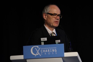
Twelve-month results from the randomised, multicentre DEFINITIVE AR pilot study suggest that there is a benefit to adding directional atherectomy in long and calcified lesions prior to using a drug-coated balloon in comparison to the use of a drug-coated balloon alone. Data from the trial were presented yesterday by Thomas Zeller, Bad Krozingen, Germany, who is a study principal investigator alongside Gunnar Tepe, Rosenheim, Germany.
DEFINITIVE AR was designed to assess the effect of treating a lesion with directional atherectomy (Medtronic/Covidien’s SilverHawk or TurboHawk plaque excision systems) followed by a paclitaxel-coated balloon (Bayer HealthCare’s peripheral paclitaxel-coated angioplasty catheter with Paccocath Technology), collectively referred to as DAART (directional atherectomy + anti-restenotic therapy), in order to generate early hypotheses for further research in this therapy area.
Claudicants (Rutherford clinical category 2–4) with 7–15cm superficial femoral and/or popliteal lesions were randomised 1:1 to either DAART (n=48) or to the paclitaxel-coated balloon alone (n=54). Patients with severely calcified lesions were assigned to a non-randomised registry arm and were treated with DAART (n=19). Baseline patient and lesion characteristics did not differ between the DAART and drug-coated balloon arms.
Technical success, defined as ≤30% residual stenosis following the protocol-defined treatment at the target lesion, was significantly higher in the DAART arm vs. the drug-coated balloon arm (89.6% vs. 64.2%, p=0.004). According to Zeller, there were significantly fewer flow-limiting dissections reported in patients treated with DAART vs. drug-coated balloon alone (2% vs 19%, p=0.01). Technical success in the DAART registry group was 84.2% and incidence of flow-limiting dissections was 0%.
At 12 months, per cent stenosis in the randomised groups was 33.6±17.7 for the DAART arm vs. 36.4±17.6 for the drug-coated balloon arm. Duplex ultrasound patency (PSVR ≤2.4, without target lesion revascularisation) was 93.4% for the DAART arm and 89.6% for the drug-coated balloon arm. Angiographic patency (≤50% stenosis and without target lesion revascularisation) was 82.4% in the DAART arm and 71.8% in the drug-coated balloon arm. In the DAART cohort lesions with a residual stenosis <30% post atherectomy did show a trend towards better angiographic patency at one year (94.1% vs. 68.8%).
“This rigorously performed pilot study suggests an added benefit for DAART over drug-coated balloons alone in long and calcified lesions. Further investigation in larger, prospective, statistically-powered randomised trials is warranted. Patients will be followed out to 24 months to assess durability of the results,” Zeller said.
Cordis launches Outback Elite re-entry catheter at CX
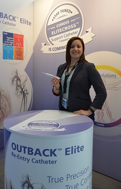
At this year’s Charing Cross Symposium, Cordis is launching the Outback Elite re-entry catheter, an enhanced version of the Outback re-entry catheter that received the CE mark earlier this week. According to the company, Outback Elite provides more control and precision and includes additional features to enable re-entry into the most complex lesions whilst facilitating positioning and delivery.
Yesterday, Thomas Zeller, Bad Krozingen, Germany, a user of the Outback re-entry catheter, spoke to CX Daily News about his experience with the device and his expectations about this new iteration.
Amongst the new features of Outback Elite are single-handed torque and deployment—the torque control is located closer to the needle actuator and the longer handle provides a more efficient slider mechanism—a robust nitinol cannula for re-entry in very complex lesions and a lubricious hydrophilic coating to help delivery.
In addition, the Outback Elite is available in a 120cm shaft length as the previous catheter but is also being launched with an 80cm shaft length indicated for ipsilateral antegrade or iliac procedures. The new size offers the advantage of having less shaft length to handle outside of the patient, increasing precision on the re-entry site.
The Outback Elite follows the successful performance of the Outback catheter. In a randomised controlled trial by Roberto Gandini et al comparing re-entry with Outback vs. manual re-entry in chronic total occlusions in the superficial femoral artery, the device achieved 100% re-entry rate vs. 42.3% with the manual re-entry technique. It also showed shorter procedure and fluoroscopy times.
Kristen Rock, global product director at Cordis, told CX Daily News, “Outback Elite’s ergonomic handle and added shaft sizes provide physicians with more control, meaning they can easily pinpoint exactly where they want the re-entry site to be.”
The Outback re-entry device will be used in a live case today at the CX Live Peripheral Arterial Cases – SFA and popliteal in the Grey Learning Centre.
CX 2015 audience voting favours standalone use of drug-coated balloons only for short SFA lesions
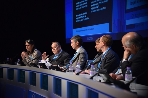
Drug-coated balloon technology is unlikely to be a successful “stand alone” treatment for the majority of medium and long length superficial femoral artery lesions, the voting results from CX2015 implied. The voting showed that the audience felt that drug-coated balloon use was best suited to short lesions. The randomised controlled trial data for drug-coated balloons are mostly based on short lesions and 57% of the audience voted “no” to the question, “Is drug-eluting balloon technology likely to be a successful standalone treatment for the majority of medium and long length superficial femoral artery lesions?” The session in which the vote was held was titled “Leaving nothing behind” and also saw the presentation of the ILLUMENATE first-in-human study 24-month results. These were favourable for the Stellarex drug-coated balloon (Spectranetics), and came close on the heels of updates on the Lutonix drug-coated balloon (Bard) and the IN.PACT Admiral drug-coated balloon (Medtronic). Atherectomy as a pretreatment also received a boost.
For the first time, CX voters also found support for atherectomy, but as a pre-treatment to the use of drug-coated balloons as described in the DEFINITIVE AR trial or as a pre-treatment to the use of stents. The majority, 77%, supported the motion “pretreatment of superficial femoral artery lesions before drug-coated balloon has CX 2015 audience support.
Thomas Zeller, Bad Krozingen, Germany, explained that “Drug-coated balloons add benefit to the endovascular approach to treat femoropopliteal disease but that there are still limitations such as recoil, calcium and dissections.”
Previous research has shown that circumferential calcification is a negative predictor for successful outcome with drug-coated balloon angioplasty. “Directional atherectomy is one of the most interesting pretreatment plaque modulating or plaque removing options available, after cutting or scoring balloons have been shown not to be very effective at this. The removal of ex-centric plaque and calcified plaque serves to reduce the problem of early recoil that limits the use of drug-coated balloon angioplasty outcome,” he said. Zeller also noted that the large majority vote favouring the use of atherectomy as a pretreatment were akin to a mandate to the industry to support large-scale randomised controlled trial to prove this concept.
In previous years, atherectomy has failed to elicit CX voter backing as a definitive treatment option in the superficial femoral artery. In 2014, 57% of the CX audience voted “no” to the motion, “I would use atherectomy for some superficial femoral artery lesions”. In 2013, 68% voted against the motion “atherectomy is the answer for the superficial femoral artery technology”.
This year’s vote shows that there is increasing support for atherectomy as a pre-treatment option rather than a definitive treatment and that the technique is clearly currently in favour of being used prior to drug-coated balloon or stents in order to reduce the risk of thrombosis. A member of the audience asked whether atherectomy should be the first option for isolated popliteal lesions considering there was a desire to leave nothing behind. Zeller, responding, said that it was frequently observed that isolated popliteal lesions were focal calcified lesions and for that indication, he considered atherectomy “an excellent first option followed by angioplasty or stenting”. “However, if we are talking about evidence to support this, there is none,” he clarified.
CX delegates at the session also heard updates on the Stanza bioresorbable scaffold (480° Medical) and Shockwave Lithoplasty for calcified artery from Andrew Holden, Auckland, New Zealand.
ILLUMENATE
Stephan Duda, Berlin, Germany, presented the results of the 24-month ILLUMENATE first-in-human study. These showed that the primary patency rate (as measured by Duplex Core lab evaluation) was 80.3% at 24 months. At 12-months, the primary patency rate was 89.5%.
“The freedom from clinically-driven target lesion revascularisation rate (as determined by the clinical events committee adjudication) was 85.8% at 24 months and it was 90% at 12 months,” Duda said.
In the study, researchers evaluated Stellarex in 50 patients with 58 lesions in a cohort that required predilatation with an uncoated balloon before the inflation of the drug-coated balloon. The mean age of the cohort was 69 years. The mean age of patients was 69.0±9.3 years; Thirty four per cent of patients had diabetes and 80% had hypercholesterolemia; Eighty six per cent were Rutherford Class 3 at baseline; the mean lesion length was 7.2cm; nearly 14% had severe calcification and the mean stenosis at baseline was 75%.
The vessel patency was associated with a significant and sustained functional improvement as resulted by the walking impairment questionnaire and by the treadmill test patient subset. There were no cardiovascular deaths or amputations reported throughout the 24-month follow-up schedule in this cohort, demonstrating the high safety profile,” Duda said.
He continued: “This study demonstrates the safety and efficacy of Stellarex for the treatment of femoropopliteal disease up to two years. The primary patency of 89.5% and 80.3% match the highest benchmark of reported rates at one- and two-years respectively. There is also significant functional benefit observed with improved walking distance, observed up to two years.”
Duda told CX Daily News: “Stellarex employs a next-generation manufacturing technology and coating formulation. This combination allows for an effective dose of paclitaxel to be transferred to the treatment site using a low drug dose density of 2µg/mm2. Pre-clinical work has shown this coating results in high coating stability with limited drug loss and an effective amount of drug in the arterial wall through 28 days.”
There is a robust clinical programme for Stellarex that is actively underway and will include up to 1,300 patients in five studies, Duda concluded.
IN.PACT SFA clinical trial update
Gunnar Tepe, Rosenheim, Germany, presented the 12-month results from the pivotal randomised trial evaluating the safety and effectiveness of the IN.PACT Admiral drug-coated balloon.
“IN.PACT SFA is a rigorously designed trial that provides level I clinical evidence supporting the safety and effectiveness of the IN.PACT Admiral when used to treat lesions in the superficial femoral and proximal popliteal arteries,” Tepe said.
The trial is a prospective, randomised, international, 57-site trial comparing the outcomes of treating a lesion with the paclitaxel-coated IN.PACT Admiral vs. standard percutaneous transluminal angioplasty using an uncoated balloon.
A total of 331 patients with claudication or rest pain (Rutherford clinical category 2–4) with single de novo or non-stented restenotic lesions ≥4cm and ≤18cm or chronic total occlusions ≤10cm were randomised 2:1 to drug-coated balloon (n=220) or angioplasty (n=111). Data was independently evaluated by blinded duplex ultrasound and angiographic core labs and by a blinded clinical events committee.
Tepe said: “Baseline clinical, angiographic and lesion characteristics were well balanced between treatment groups. Mean lesion length was 8.94±4.89cm in the drug-coated balloon arm vs. 8.81±5.12cm in the angioplasty arm (p=0.815), representing the longest lesions evaluated to-date in a randomised drug-coated balloon trial.”
A total of 59.3% of lesions in the drug-coated balloon arm were calcified vs. 58.4% in the angioplasty arm (p=0.097), and 25.8% of lesions in the drug-coated balloon arm were total occlusions vs. 19.5% in the angioplasty arm (p=0.222).
“At 12 months, primary patency in the drug-coated balloon arm (freedom from clinically-driven target lesion revascularisation and freedom from restenosis as determined by duplex ultrasound PSVR≤2.4) was superior to the angioplasty arm (89.8% for the drug-coated balloon vs. 66.8% for the angioplasty arm, Kaplan-Meier analysis at day 360 showed that p<0.001). Clinically-driven target lesion revascularisation was statistically lower for the drug-coated balloon arm vs. the angioplasty arm (2.4% vs. 20.6%, p<0.001). Regarding clinical endpoints, we can conclude that patients treated with IN.PACT Admiral perform more favourably than angioplasty in terms of quality of life and walking capacity and angioplasty treated patients require 88% more interventions to achieve the same clinical outcome,” Tepe said.
“Patients are being followed out to five years post-procedure and two-year data are forthcoming,” Tepe concluded.
Lutonix update
Dierk Scheinert, Leipzig, Germany, spoke on the topic “New insights from Levant 2 randomised data” that included data from subgroup and post-hoc analysis. “Levant 2 demonstrated superior patency for Lutonix against angioplasty with a 29.4% improved patency over angioplasty. The device was effective in a challenging patient population and showed target lesion revascularisation rates of 89% that were consistent in both the clinical trial and real-world registry,” he said.
Scheinert noted that Levant 2 had demonstrated the safety of Lutonix showing low rates of re-intervention for thrombosis and embolism. “Patients who received Lutonix DCB reported clinical benefits in Levant 2 and showed sustained improvement in Rutherford class and improvement in self-reported walking distance scores” he said.
Fabrizio Fanelli, Rome, Italy, outlined the current status of drug-coated balloon use in the superficial femoral artery. “There are more than 10 drug-coated balloons that are commercially available in Europe. All of them are based on the use of the same drug, paclitaxel, but the dosage is different, varying between 2µg/mm2 and 3.5µg/mm2. Moreover, each drug-coated balloon has a different excipient and a different coating technology,” he said. Most drug-coated balloons have passed their proof-of-concept test, but few have offered high-quality clinical evidence and programmes, even fewer have delivered best-in-class outcome, he emphasised.
Fanelli highlighted that studies such as IN.PACT SFA, LEVANT-2 and ILLUMENATE FIH were all multicentre studies with results that have been validated by independent core labs and that these data were essential in evaluating the quality of evidence and the quality of outcomes. “Only the most reliable data must be taken into consideration. There is no drug-coated balloon ‘class effect’. Each drug-coated balloon is to be judged upon the existence and value of its own data,” he stated.
Drug-coated balloons are cost-effective
Michael Jaff, Boston, USA, told CX 2015 delegates that while there are no prospective data collection studies, two publications have demonstrated that drug-coated balloons are a cost-effective alternative to angioplasty.
Jaff said: “Using current modelling, drug-coated balloons cost the system less than angioplasty, bare metal stents and drug-eluting stents, due to the lower target lesion revascularisation rates. It is critical that future randomised trials of one revascularisation technology include direct measurements of cost of care.”
He told delegates that the future was to be all about economic value and that in the previous week, the US payer system was in the process of reviewing the cost, efficacy and outcomes of all peripheral artery interventions for patients with intermittent claudication.
Jan B Pietzch and colleagues, publishing in Catheterization and Cardiovascular Interventions (2014), in studying the impact on payers and providers of the four main endovascular strategies for the treatment of infrainguinal peripheral artery disease in the USA and Germany, showed that drug-coated balloons and drug-eluting stents, compared to bare metal stents and percutaneous transluminal angioplasty, are associated with lower probabilities of target lesion revascularisation and therefore, cost savings for payers. Another publication, from BC Kearns and colleagues, published in the British Journal of Surgery in 2013, that set out to perform an economic evaluation of the cost-effectiveness of endovascular enhancements to percutaneous transluminal balloon angioplasty with bail-out bare metal stents for infrainguinal peripheral arterial disease, found that the use of drug-coated balloons represents a cost-effective alternative to the use of angioplasty with bail-out bare metal stents.
Jaff made the point that comparing the cost of using drug-coated balloons with the costs of percutaneous transluminal angioplasty, bare metal stents and drug-eluting stents was a “complex proposition”.
“Device costs are only one part of the equation. If the procedure offers greater durability, as defined by reduction in clinically driven target lesion revascularisation, the cost of care for that patient with peripheral artery disease will be lower than a comparable device requiring repeat interventions over a defined period of time,” Jaff concluded.
Direct balloon angioplasty
The panel agreed that more data were needed on whether pre-dilatation was needed before drug-coated balloon angioplasty was performed or not and called for a trial to compare the outcomes achieved with drug-coated balloon use after pre-dilatation to those achieved with direct angioplasty with drug-coated balloon.
Leaving something in
Speaking on the paradigms and importance of lesion lengths and types in the afternoon session on increasing lesion length options and various stent controversies, William Gray, New York, USA, made the point that lesion length is the only clearly identified driver of long-term outcomes. He drew attention to the fact that “Short-term outcomes may also be affected with increasing stent usage with increasing lesion length. Chronic total occlusion and diabetes do not appear to effect outcomes. The calcification grading is problematic, but likely has implications for both short- and long-term outcomes and adjunctive/specialised therapies may mitigate this effect.
Holden noted that five years ago lesion length had had a direct impact on patency with the technology that was available then. “Now with an armamentarium that consists of drug-coated balloons, biomimetic stents, drug-eluting stents, covered stents and atherectomy, we are saying that that paradigm is changing. Are we seeing less impact of lesion length on patency?” he asked. Gray noted that in the main, lesion length was the main driver of patency but that newer technologies and adjunctive therapies could modify the longer lesions in the drug-coated era and that he remained optimistic about this development.
Edited live aortic and carotid cases presented at CX for the first time
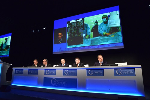
CX’s new Edited Live Case session, which aims to highlight the technical aspects of the aortic and carotid main programme, was launched yesterday with a selection of cases exploring how to apply different techniques in complex situations to achieve the optimum result for the patient.
Roadsaver – Carotid stenting with micromesh
The Roadsaver (Terumo), a self-expanding stent that has a dual layer micromesh design for sustained embolic protection, was used to treat a 73-year-old female with a high-grade symptomatic lesion in the internal carotid artery, Arne Schwindt (Munster, Germany) reported. He explained that she had an ipsilateral Amaurosis fugax eight weeks’ prior to the intervention and computed tomography (CT) angiography and Duplex confirmed a 80% right internal carotid artery stenosis. The patient also had arterial hypertension, hypercholesterolaemia, and type II diabetes.
Further to a having dual mesh design, Schwindt commented, the Roadsaver has “a flexible weave completely made of nitinol, an extremely flexible delivery system, and is retrievable and repositionable”. He explained that although accessing the lesion was the key challenge in the case, the risk of dislodging the stent’s guidewire system was low because of the system’s flexible delivery system.
According to the six-month follow-up result, the patient had no neurological events during the peri or post procedural period. Furthermore, convergent colour Doppler showed that the stent was patent with vmax of <100cm/seconds (previously, it was 190cm/sec). Schwindt concluded that the stent has “proven good deliverability, wall adaptation, and can be used with a variety of protection devices in challenging anatomies and lesions.”
Relay – Scalloped thoracic graft to the left subclavian artery and branched arch procedure
Mo Hamady and Joost van Herwaarden (London, UK, and Utretch, The Netherlands, respectively) jointly presented the case of a 43-year-old female patient with known hypertension who arrived at the hospital after she fell from steps at home. Because the patient presented with back pain, CT angiography was performed and this showed a non-traumatic saccular aneurysm of the thoracic aorta (ie. ductus diverticulum).
The presenters explained that the 9.5cm type 1 thoracic aortic aneurysm involved type III aortic arch, starting from the level of the innominate artery and extending to the distal thoracic aorta. There were more than 90 degree double bends at the distal end of the aneurysm.
According to van Herwaarden, the distance from the left subclavian artery to the aneurysm was too short for thoracic endovascular aortic repair (TEVAR) below the subclavian artery, and “further the ‘young’, highly curved aortic arch might have caused bird-beaking of a stent graft deployed in the aortic arch”.
Treatment options were TEVAR with covering of the left subclavian artery with or without revascularisation, TEVAR using a custom-made stent graft with a scallop for the left subclavian artery, or open repair.
The Relay (Bolton Medical) device, a custom-made branched thoracic stent graft with proximal scallop, was deployed in the aorta. Bridging stents to the innominate and left common carotid arteries were used to seal the aneurysm. The left common carotid to left subclavian artery bypass was used to revascularise the covered native subclavian artery.
Technical success was achieved and a postoperative CT angiography at one month showed exclusion of the aneurysm with open left subclavian artery and without bird-beak configuration.
Treatment of a chronic type B aortic dissection with Valiant Captivia after ascending aorta and arch surgery
Luigi Lovato (Bologna, Italy) presented the case of a 73-year-old female with a history of hypertension, ascending aorta surgery for aneurysm and chronic type B dissection from middle descending thoracic aorta. The patient was submitted to arch aneurysm surgical replacement. Serial CT imaging showed progressive severe dilation (>5mm/year) of aortic isthmus and middle thoracic aorta false lumen, joining 5.5cm at the level of the big entry tear with appearance of partial false lumen thrombosis.
The patient was considered for TEVAR of both aortic lesions. Lovato said that challenges arose due to the surgical reconstruction anatomy with angulation between the prosthesis and native aneurysmatic aorta and difficult visualisation of the reimplanted epiaortic vessel, creating a relatively short proximal neck, and discrepancy between the proximal and distal neck, because the sinusoidal intimal flap and the relatively distal entry tear forced them to cover the whole thoracic aorta extending next to the coeliac trunk where the true lumen is small.
He said that the Valiant Captivia (Medtronic) stent graft was selected because “it combines a solid capture tip system to obtain a high controlled stent deployment and low risk of wind-sock and bear-beck effect and, at the same time, an optimal hydrophilic delivery system to manage the advancement through a relatively small femoro-iliac access and tortuous aorta”.
During the procedure, Lovato explained, they adopted a different approach, delivering the distal stent graft first to solve the neck discrepancy. The second stent was deployed afterwards, proximally, in the surgical prosthesis and with the distal part inside the first distal stent. The sizing was sorted to achieve the best oversizing in the prosthesis, avoiding type I/III endoleak.
The pre-discharge CT control (six days later) showed an optimal stent graft positioning without type I/III endoleak. A small type II endoleak was visible at isthmic level. There was a moderate retrograde flow from the abdominal false lumen reaching the middle thoracic aorta, but only in the late CT scan.
“The case demonstrates the need for careful planning to meet the anatomical challenges in patients with previous arch surgical reconstructions and the feasibility and safety of this ‘distal first approach’ of TEVAR for type B dissection. This technique can solve the frequent neck discrepancy of these patients avoiding the severe complication of distal false lumen aneurysmal degeneration, caused by the intimal flap rupture induced by the stent graft radial forces,” Lovato concluded.
ZENITH Alpha thoracic endograft
Geert Schurink (Maastricht, The Netherlands) reviewed the case of an 80-year-old female whose thoracic aneurysm was discovered after she underwent a computed tomography (CT) scan for a deep vein thrombosis. He said she was “a very fit” 80-year-old but did have a kyphosis and moderate femoral pulses.
The patient underwent TEVAR with the Zenith Alpha thoracic endograft (Cook Medical). Afterwards, in the recovery unit, mean arterial pressure was >85mmHg and a cerebral spinal fluid drain was not put in because of her concerns about her kyphosis. However, two days after the procedure, she lost sensation in both lower legs and experienced weakness in her left leg. A cerebral spinal fluid drain was put in and her mean arterial pressure was >90mmHg. Schurink reported that the patient made an “almost complete recovery”. He added that he did not believe the loss of sensation was a result of an embolization (in the distal aorta) because if it had been, he said, she would not have made a recovery. Furthermore, there were no signs of emboli on angiography.
After the case was presented, the audience not only discussed when to put in a cerebral spinal drain after TEVAR but also reviewed whether patients were allowed to get too hypotensive compared with their relative baseline pressure.
Nellix – Endovascular aneurysm sealing (EVAS) with polymer in the aortic sac
In the case that Michel Reijnen (Arnhem, The Netherlands) reported, a 70-year-old male patient, with a cardiopulmonary history, presented with an infrarenal aneurysm with a maximum diameter of 55mm.
The patient was treated using the Nellix endovascular aneurysm sealing system (Endologix). Reijnen explained that Nellix consists of two balloon-expandable endoframes surrounded by endobags that are filled in situ with a biostable polyethylene glycol (PEG)-based polymer to completely fill the aneurysm sac, thus sealing the entire aneurysm.
The case showed that the infrarenal neck diameter and length were respectively 28mm and 35mm. Access was adequate and the calculated volume to seal the aneurysm was 64ml. After a bilateral cutdown, an angiography was performed with a calibrated catheter. Subsequently, two Nellix endosystems were inserted, positioned and deployed. A prefill with saline solution, to a pressure of 180mmHg, was performed showing a complete exclusion of the aneurysm. The saline solution was then replaced by 55mL of polymer to reach a final pressure of 190mmHg. During curing the Nellix balloons were re-inflated.
Completion angiography showed a complete exclusion of the aneurysm with patent renal and hypogastric arteries. Throughout one year follow-up, there were no complications.
Reijnen advised that the “key points for success are case planning and extensive knowledge of the device and technique, which differs from regular EVAR”.
The INCRAFT low profile device introduced percutaneously
A patient with a 55mm aortic abdominal aneurysm was the subject of the case that Hany Zayed, Gianbattista Parlani and Jost Philipp Schäfer (London, UK; Perugia, Italy; and Kiel, Germany, respectively) presented. They reported on how the patient was treated with an Incraft endograft (Cordis).
The procedure was performed under local anaesthesia with a bilateral percutaneous approach. The presented case showed the endograft’s main body delivery and the possibility to reposition the graft, as well as the Iliac limbs implantation accommodating the device into the broad spectrum of maximum and minimum overlapping in order to obtain the correct length of the device to preserve the hypogastric artery.
The completion angiography performed at the end of the procedure showed precise endograft placement with the start of the covered portion of the device immediately below the lowest renal artery and total coverage of both common iliac arteries with preservation of the hypogastric artery.
Parlani noted that the key message of the presentation “is that this ultra-low profile device is designed for proximal and distal placement accuracy and allows for customisation during the procedure to accommodate a wide range of anatomical sizes. This broad anatomical coverage is offered with a minimal number of product codes for easier pre-procedural planning”.
Challenging AAA anatomy treated by Endurant IIs stent graft
Medtronic’s Endurant IIs stent graft has several advantages and benefits for managing patients with challenging anatomy, Johannes Gahlen (Ludwigsburg, Germany) said in his case presentation of a 64-year-old male with an abdominal aortic aneurysm of 5.2cm.
He explained that the device has expanded anatomical customised options, including a 20% reduction in distal diameter compared with select Endurant II bifurcation stent grafts, in-situ sizing with select ipsiliateral limbs, allowing a 3–5 stent overlap for adjustment curing cases, and easier pre-case planning to simplify sizing. It has also has a “better anatomical fit” with “less material encumbrance in tight aortic bifurcations and “better precision in distal landing to avoid type IIb endoleak or unintended coverage of the hypogastric artery.
Gahlen reported that, in the case he was presenting, endovascular aortic aneurysm repair was performed with the graft using the three-piece approach. He described the patient’s anatomy as challenging because there was kinking and stenosis in the left common iliac artery and the right common iliac artery was dilated.
According to Gahlen, the usual practice is to perform the follow-up CT scan at six months, but three-month follow-up was planned for this patient because the device was relatively new.
The Aorfix endovascular stent graft
The patient in the case presentation from Robert Beasley (Miami, USA) was an 81-year-old male with a history of diabetes, hypertension, chronic obstructive pulmonary disease, coronary artery disease, and smoking (he quit in 2009). Beasley commented that the patient was found to have a 7.8 x 7.3 x 10cm abdominal aortic aneurysm with an angle of
He explained that the patient was treated with an Aorfix endovascular stent graft (Lombard). Features of this device include, according to Beasley, radioplaque markers on the stent body graft and legs for “precise positioning”, 8mm sealing zones at proximal distal ends of stent grafts, and a “fishmouth” fixation mechanism (the proximal end of the graft expands into a fishmouth shape). Beasley called this mechanism a “unique design concept” that allows the endograft to be placed transrenally and has four pairs of hooks to “resist migration forces”.
The E-liac device in complex iliac aneurysm
In his presentation, Jost Philipp Schaefer (Kiel, Germany) explained that a 56-year-old male patient developed a dissected iliac aneurysm in the left common iliac artery after undergoing surgical repair of an aortoiliac aneurysm with an aortofemoral (right) and aortoiliac (left) graft interposition.
The dissection caused a severe stenosis at the ostium of the internal iliac artery with symptoms of buttock claudication and erectile dysfunction. Via combined left groin and transbrachial approaches, the dissected iliac aneurysm is repaired by utilising the E-liac device and an E-ventus stent graft (Jotec) as bridging stent. The applicable techniques were demonstrated step-by-step, including the completely percutaneous approach under local anaesthesia, the successive deployment of the iliac-side-branch graft, and the placement of the bridging stent graft.
Schäfer said that this Edited Live Case “demonstrates the technique of endovascular repair of iliac artery aneurysm with the new E-liac device. Available off-the-shelf, the E-liac device is a low-profile iliac-side-branch graft, offering a percutaneous treatment option for a wide range of aortoiliac aneurysmatic pathologies”.
Control of infection with prosthetic grafts: added value of INTEGARD SYNERGY
Jean-Paul de Vries (Nieuwegein, The Netherlands), in his case presentation, explained how the Intergard Synergy (Maquet) graft was used to treat a 62-year-old female with (as identified on CT scan) a mycotic abdominal aorta combined with Leriche syndrome. He explained that the graft was the only one to be loaded with two different antiseptics, adding that these antiseptics (silver acetate and triclosan) are “not limited to one target and do not have the likelihood to produce bacterial resistance”. De Vries added that the graft was “designed for routine prophylactic use” and was not associated with any contraindications for contaminated implant sites.
In this case, the patient was on the hospital ward for 12 days after the operation and received penicillin for eight weeks afterwards. According to de Vries, there was no sign of infection at the two month postoperative CT scan.
Hello world!
Welcome to WordPress. This is your first post. Edit or delete it, then start blogging!
“Ask the expert” workshop dedicated to imaging
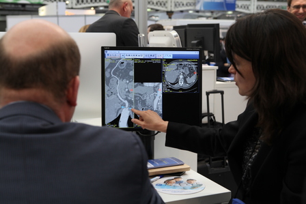
To complement the CX Imaging Day morning session, in the afternoon delegates will have the opportunity to learn about new imaging systems in a hands-on workshop including different imaging modalities used in vascular and endovascular management.
The “Ask the expert” workshop will be held at the GE Healthcare, Hansen Medical, Philips Healthcare and Siemens imaging stands in the Exhibition Hall. Physician-experts in imaging will be giving demonstrations combined with talks focusing on sizing, 2D/3D fusion imaging and how to achieve optimal dose levels.
Imaging industry stands programmes
The programmes at the imaging companies’ stands will combine hands-on demonstrations and talks. They will run from 1:30pm until 5:30pm on Thursday 30 April.
GE Healthcare booth 130
Faculty/demonstrators: Stéphan Haulon, Lille, France and Adrien Hertault, Lille, France
- EVAR procedures: From sizing to fusion
- Achieving optimal dose levels
Hansen Medical booth 221
Programme to be announced
Philips Healthcare booth 318
Faculty/demonstrators: Frank Vermassen, Gent, Belgium and Jim Reekers, Amsterdam, Netherlands
- Vessel Navigator: Learn all about the latest innovation in Live Image Guidance solutions
- 2D Perfusion: Get insight in the latest perfusion angiography solution
- Veradius Unity: Experience the innovative vascular workflow of our newest Mobile C-arm with flat detector
- How to build a Hybrid room: Considerations when purchasing a Hybrid room
Siemens booth 420
Faculty/demonstrators: Dittmar Böckler, Heidelberg, Germany (second speaker TBA)
- Adding smooth to smart in EVAR guidance
- Learn about innovative tools to treat complex endovascular aortic repair cases with confidence
- The new PURE Software for the Artis zeego enables a smooth workflow to use the innovative smart tools at the day of operation