


CX NEWS
Leave nothing behind: Bioresorbable devices are an “exciting prospect” for the future
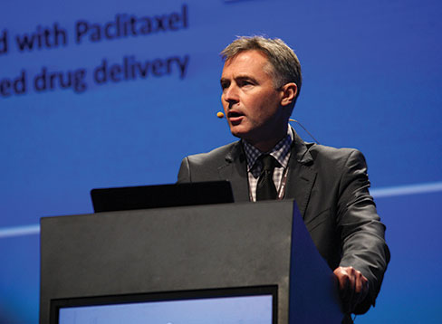
Andrew Holden, Auckland, New Zealand, presented data for on the developments in bioresorbable technology yesterday. He said that, as drug-eluting balloon data are limited to short and intermediate length lesions, bioresorbable stents or scaffolds are an “exciting prospect” for the future.
He said that the long and mobile femoropopliteal arterial segment is a challenging environment for endovascular intervention; for more complex lesions a scaffold is often required to prevent residual stenosis and flow-limiting dissection. An anti-restenosis strategy is also important, particularly in claudicants where long-term patency is vital, he noted.
“Drug-eluting balloons are highly promising but have only been studied for any duration in relatively short lesions. Drug-eluting stents have shown satisfactory patencies in intermediate length lesions but suffer the problems of a permanent self-expanding metallic implant.”
Addressing this problem with drug-eluting stents, Holden said for many years bioresorbable stents have been eagerly awaited in the superficial femoral artery.
“A bioresorbable stent may provide a scaffold to optimise the acute result after angioplasty without the long-term irritation of a self-expanding stent. Such a scaffold must withstand the hostile environment of the superficial femoral artery, provide mechanical support and integrity through vessel healing, remain biocompatible through resorption and facilitate drug delivery,” he said.
Three bioresorbable stents have recently been studied in the superficial femoral artery. The Abbott Esprit 1 trial used a balloon expandable PLLA scaffold in iliac and femoral arterial lesions ≤50mm in length that could be treated by a single 6.0mmx58mm device. The study reported excellent procedural success. Although lesion length was short (mean 35.7mm), the patency data and improvement in Rutherford-Becker status at one year was very encouraging, Holden said.
The 480 Biomedical Stanza stent has been studied in the STANCE trial. This flexible, self-expanding stent is a PLGA and bioreorbable elastomer composite and fully resorbs in 12–15 months, according to Holden. Acute performance and subsequent stent strut encapsulation and resorption has been evaluated with optical coherence tomography (OCT). This study reported excellent procedural success and acute stent performance, treating longer lesions (up to 90mm).
Holden explained that late lumen loss seen in the first cohort of patients was due to a combination of vessel recoil and neointimal hyperplasia. The device was modified in a second patient cohort with minimal vessel recoil. A paclitaxel, drug-eluting version of the scaffold has recently entered the clinic in the SPRINT trial, he added.
Holden also reported that the Igaki-Tamai bioresorbable scaffold (Igaki Medical Planning) has been used in the superficial femoral artery in a 30 patient cohort. Acute procedural results were very good, as in the STANCE trial.
“Binary restenosis rates at 12 months were unacceptably high and further modifications are planned. Small clinician initiated trials using coronary bioresorbable stents in the tibial arteries are being performed but meaningful results are not yet available,” Holden commented.
“There has been considerable progress with bioresorbable stents in the superficial femoral artery. The technology is not yet ready for routine clinical practice but that exciting prospect should not be far away,” Holden concluded.
“New approach to superficial femoral artery reconstruction is highly versatile”
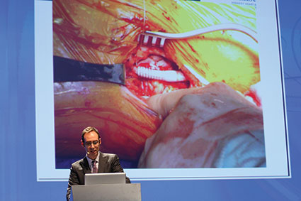
In his technical report of the Hybrid Vascular Graft (Gore) for the reconstruction of the superficial femoral artery, Jean Bismuth (Houston, USA) said the graft allowed a rapid sutureless anastomosis and was highly versatile in complex situations.
Bismuth reported that the graft is the first device that has been designed to “address the ever expanding number of hybrid operations, which combine both endovascular and open surgical techniques.” He explained that it was an expanded PTFE vascular prosthesis that had a constrained nitinol section and added that it “greatly expanded” the treatment options for dialysis access, aortic debranching, and arterial bypass procedures. Bismuth commented: “The graft allows the surgeon to minimise the invasiveness of a procedure all the while benefiting from the advantages of a bypass.”
He explained that although initially the graft was launched for arteriovenous dialysis access, it now had the same indications as “any vascular graft”. For example, at his centre, where the graft has been used in 150 cases, it has been used for “a good proportion of iliofemoral bypasses and a bunch of visceral debranching as well.”
Bismuth commented: “In the femoropopliteal segment, the Hybrid graft permits access through a smaller incision to an artery which is either hard to reach behind the knee or is diseased. The nitinol reinforced segment of the graft can be introduced through a less invasive access and can take advantage of the stent to treat a stenotic segment.”
As well as reviewing the advantages of the graft, Bismuth also discussed the “pearls for success” with the device and these included the fact that “imaging was key” to successfully accessing patient anatomy—“you have to know what you are going into”, he noted. Another pearl was that correct sizing was “essential” as he said that the graft should not be oversized by more than 20%. Bismuth added that he never oversized by more than 1mm and that correct sizing could mean that sutures were not needed at all. He explained: “Gore recommends a couple of patching sutures, but I have never put any in. It is a personal choice and I have never had an issue with needing to use sutures. It is all about the sizing. If you only upsize by 1mm, you are probably not going to have any problems.” Other tips for success included not completely inserting the nitinol reinforced segment into the artery, ensuring adequate outflow and inflow, and covering all of the disease in the artery.
Bismuth concluded: “Hybrid procedures are likely to be more prevalent; the Hybrid graft allows a rapid sutureless anastomosis, particularly for difficult sites and rapid bailout. It is highly versatile in complex cases.”
BASIL 2 randomised trial launched to address need for data for endovascular interventions for severe limb ischaemia

Andrew Bradbury (Birmingham, UK) reported that a new randomised controlled trial, BASIL 2, has been launched because there is an “urgent need” to undertake pragmatic, scientifically robust and publically-funded randomised controlled trials of endovascular interventions in patients with severe limb ischaemia.
Bradbury stated that the findings of BASIL 1, which showed a significant improvement in overall survival with endovascular therapy in patients with severe limb ischaemia compared with surgery at 7.3 months (but not at two years), and those of other studies have been used to justify “an endovascular approach” in this group of patients. However, he added that BASIL 1 may no longer be relevant because of several changes in the treatment of severe limb ischaemia since the study was published. For example, Bradbury reported, endovascular therapy had “changed beyond recognition”, interventional radiologists were more skilled at performing endovascular therapy, and surgeons could now perform hybrid procedures. Furthermore, other studies in this area were industry sponsored and had not produced the answers needed to make nation-wide decisions about which therapy to use. Bradbury said, during development of guidelines for peripheral artery disease, the UK’s National Institute for Health and Care Excellence (NICE) were “shocked” that the day-to-day decisions for the management of severe limb ischaemia were being based on such a “lack of evidence”.
Therefore, the BASIL 2 has been launched to provide more data on the effects of endovascular therapy compared with surgery in patients with severe limb ischaemia. Bradbury reported that in the superiority trial, 600 patients with below-the-knee or femoropopliteal atherosclerosis will be randomised to receive “best endovascular therapy” or a vein graft. He added that the recruitment process, which was due to start in a few weeks, would last for 36 months and the primary endpoint was the rate of amputation-free survival at 33 months. To show superiority of endovascular treatment, Bradbury reported, there needs to be a 15% difference in the primary endpoint between the groups.
The study will be funded by the National Institute for Health Research and, according to Bradbury, has received “overwhelming support” from both interventional radiologists and vascular surgeons. He acknowledged that recruitment for BASIL 1 had been difficult—“it is the reason why my hair is grey; in fact, it is a wonder that I have any hair at all”—but said the vascular community were now more accepting of the need for randomised controlled trials.
Concluding, Bradbury said that there was an “urgent need” to undertake pragmatic, scientifically robust and publically-funded randomised controlled trial of endovascular therapies in severe limb ischaemia that were powered for clinically important endpoints and include a full cost-effectiveness analysis.” He added that the BASIL 2 would look at the health costs and, for “the first time”, the social costs of the interventions in the study. “Without data from such randomised controlled trials, we cannot be sure that endovascular interventions are not going to be associated with net harm or suboptimal use of precious health resources,” Bradbury said.
Practical issues arising from hyper-acute carotid interventions

In Leicester, UK, rapid-access surgery for symptomatic carotid stenosis has been offered since October 2008. Ross Naylor reviews the practical lessons that have been learned with the experience. He discussed this topic at CX35 on Tuesday.
By Ross Naylor
When the trials randomised “recently symptomatic” patients to carotid endarterectomy or medical therapy, symptoms had to have occurred
Consequently, there has been a move towards performing surgery as soon as possible after the index event (ie treating transient ischaemic attack on a par with unstable angina). NICE advise that patients should undergo surgery
Some surgical/interventional colleagues have not actively embraced the move towards expedited intervention. For them, patients benefit from a period of stabilisation and assessment (in order to reduce procedural risk), while others believe that intervening early is associated with an unacceptably high procedural risk that may negate any benefit conferred through early intervention. The reality is, however, that the surgeon who operates within two weeks with a 10% procedural risk is still likely to prevent more strokes (in the long term) than the surgeon who defers surgery for four weeks and then operates with a 0% risk!
The Leicester Unit has offered a rapid-access surgery service since October 2008. All patients are seen in a 24/7 cerebrovascular clinic and those with 50–99% stenoses are transferred to the Vascular Unit for expedited surgery. 400+ symptomatic patients have now been treated (12%
First; it is not unusual (in the hyper-acute setting) for the duplex operator to comment that there is a critical stenosis that does not appear to open out into a normal calibre vessel. In the past (when patients were randomised some time after the index event), this might have been diagnosed as “near occlusion” (little benefit from surgery), but this is not the case in the hyper-acute setting. The diagnostic “give-away” is that high velocities are maintained across the stenosis (even if the distal lumen cannot be visualised) and CT angiography almost always shows a reconstructable vessel. In “near occlusion”, there are very low systolic velicities and little or no diastolic flow. Accordingly, corroborative imaging is mandatory (in the hyper-acute period) before recommending against urgent carotid surgery.
Second; anyone setting up this kind of service better get used to seeing recurrent events in up to 15% of patients between admission and surgery (despite being on antiplatelet and statin therapy). Up to 40% of patients referred acutely will have spontaneous embolisation on transcranial Doppler. Recurrent events (prior to surgery) were rarely encountered in the past, largely because patients were not referred so quickly.
Third; patients undergoing surgery (using general anaesthetic) with a pre-existing neurological deficit will almost always suffer a transient worsening of their deficit postoperatively. In the past, this was an indication for re-exploration. The “key” to management is how quickly the patient recovers from anaesthesia. If it is relatively quick, they will return to their pre-operative neurological status within an hour or so. Accordingly, it is important to warn recovery staff who may otherwise be alarmed at the apparent neurological deterioration.
Fourth; be prepared to adopt even more obsessive attention to surgical technique. We have found that careless skin preparation can trigger embolisation and transcranial Doppler is invaluable in warning of the embolising (unstable) patient during carotid mobilisation.
Fifth; be prepared to encounter a higher prevalence of post-endarterectomy hypertension (25% in theatre recovery, 25% back on the ward). This is usually seen in patients with poorly controlled blood pressure pre-operatively and it is essential that medical/nursing staff have guidelines for managing this condition. If you are going to treat patients in the hyper-acute period, you cannot adopt an ad hoc approach to blood pressure management, as patients will be subject to a greater risk of hyperperfusion stroke or intracranial haemorrhage.
Finally; (and contrary to what has previously been expected), our experience of operating in the hyperacute period has not been associated with a significant increase in procedural risk.
However; if you still harbour doubts about the benefit of intervening in the hyperacute period, ask yourself how you would want to be treated should you suffer a transient ischaemic attack and have a significant carotid stenosis?
Thought so! Don’t your patients deserve the same?
Diffusion-weight magnetic resonance imaging in carotid artery interventions

Following carotid intervention, the number of detectable diffusion-weight magnetic resonance imaging (DW-MRI) lesions is an order of magnitude greater than adverse clinical event (stroke/death). This lends credence to the use of DW-MRI as a surrogate endpoint allowing comparisons of interventional strategies in studies with reduced sample size, wrote Sumaira Macdonald, Newcastle, UK. She discussed this topic at CX35 on Tuesday 9 April.
By Sumaira Macdonald
The International Carotid Stenting Study (ICSS) sub-study comparing DW-MRI lesions in patients undergoing largely filter-protected carotid stenting and carotid endarterectomy demonstrated significantly fewer DW-MRI lesions after carotid endarterectomy, implying superior of control of procedural microemboli. Sixty-two of 124 (50%) patients undergoing distal filter-protected transfemoral carotid artery stenting and 18 of 107 patients undergoing carotid endarterectomy (17%) had new DW-MRI lesions (p<0.0001). Individual lesions were smaller in the carotid artery stenting group than in the carotid endarterectomy group (p<0.0001). Of the DW-MRI positive scans following carotid artery stenting, 25 (34%) resulted from unprotected carotid artery stenting and 37 (75%) resulted from filter-protected carotid artery stenting (p<0.019). Total lesion volume per patient did not differ significantly between patients undergoing carotid artery stenting and those undergoing carotid endarterectomy.
Two small randomised trials compared proximal embolic protection (Medtronic MoMa) with distal filters during carotid artery stenting. There were substantial or significant reductions in DW-MRI lesions for the MoMa compared with filter protection.
There were significantly fewer DW-MRI lesions in the MoMa group ipsilateral to the carotid lesion (p<0.0002) but no difference in the DW-MRI lesions in the contralateral hemisphere, implying the embolic penalty associated with catheterisation of the arch/great vessel origins for transfemoral carotid artery stenting. There was also a significant difference in favour of the MoMa system for lesion volume 0 [0 to 0.84] vs. 0.47 [0 to 2.4cm3] (p<0.0001).
The PROOF first-in-man analysis of high flow rate flow reversal via direct common carotid artery access (MICHI System) evaluated 65 patients, 48 of who had pre- and post-carotid artery stenting DW-MRI read by two independent US neuroradiologists. Eight of 48 patients had new DW-MRI lesions (16.7%).
Another study examined patients undergoing transcervical carotid artery stenting with flow reversal or distal filter-protected transfemoral carotid artery stenting. DW-MRI lesions were found in four of 64 transcervical (12.9%) and in 11 transfemoral (33.3%) patients (p=0.03). In multivariate analysis, age (relative risk, 1.022; p<.001), symptomatic status (relative risk, 4.109; p<.001), and open-cell vs. closed-cell stent design (relative risk, 2.01; p<.001) were associated with a higher risk of lesions in the transfemoral group but not in the transcervical group.
The low rates of DW-MRI lesions in studies of carotid artery stenting with flow reversal via direct carotid access are commensurate with carotid endarterectomy, presumably resulting from more effective embolic control and avoidance of catheterisation of the arch.
A prospective study of 110 patients undergoing filter-protected transfemoral carotid artery stenting investigated the fate of silent DW-MRI lesions. Twelve of 30 DWI lesions persisted, resulting in a lesion reversibility rate of 60%. Seventy-five per cent (12/16) of the cortical lesions disappeared while only 30% (3/10) of subcortical lesions disappeared. Eighty-three per cent (14/17) of lesions measuring 0–5mm disappeared while only 31% (4/13) of lesions measuring >5mm disappeared. It was concluded that a large number of silent ischaemic lesions visualised on the DWI images post-carotid artery stenting disappear within months and therefore the extent of permanent carotid artery stenting-related cerebral damage may be overestimated.
The most recent analyses of the ICSS sub-study data set revealed that patients in the carotid artery stenting group had more acute (relative risk 8.8, 95% CI 4.4-17.5, p<0.001) and persisting lesions (relative risk 4.2, 1.6-11.1; p=0.005) than patients in the carotid endarterectomy group. However, the rate of conversion from acute to persisting lesions was lower in the carotid artery stenting group than in the carotid endarterectomy group (RR 0.4, 0.2-0.8; p=0.007).
Systematic reviews have failed to provide consistent data across included studies comparing cognitive outcomes following carotid artery stenting and carotid endarterectomy.
Of 1,713 patients included in the ICSS, 140 of 177 patients enrolled in two Dutch centres had neuropsychometric testing at baseline and 120 at follow-up. Ten domains were examined, including executive function. There were no significant difference in overall cognition between patients undergoing carotid artery stenting and carotid endarterectomy despite the impressive difference in DW-MRI lesions counts between carotid artery stenting and carotid endarterectomy.
“Standard” filter-protected transfemoral carotid artery stenting generates more DW-MRI lesions than carotid endarterectomy but technical modifications (proximal embolic protection, direct carotid access) allow carotid artery stenting to more effectively compete where microemboli are concerned. Clinical correlation, with regards cognitive function is poor, implying either that a large number of DWI lesions are clinically irrelevant or that neuropsychometry is a blunt tool. DW-MRI is a reasonable secondary endpoint for carotid interventions, but without watertight clinical inference, the use of DW-MRI as a primary endpoint remains an unproven convenience.
CX Non-Cardiovascular Imaging Day embraces other specialties
On Tuesday 9 April, in the CX Non-Cardiovascular Adavanced Imaging Day session, Roger Greenhalgh, London, UK, introduced the speakers from the respective companies who spoke about building a hybrid operating room, the financial implications of doing so, and laser-guided hybrid suites.
In the afternoon session, chaired by John Primrose, president of the association of Surgeons of Great Britain and Ireland, showed delegates on the following topics:
- Florian Gebhard, Ulm, Germany—Making the hybrid operating room cost effective: the multidisciplinary hybrid operating room, and improving trauma care
- Manuel Ritter, Mannheirn, Germany— Radiation safety: management of dose in the hybrid operating room, and improvement of PCNL by Uro-Dyna computed tomography (CT)
- Helmut Isringhaus, Völkinghaus, Germany—Minimally-invasive resection of small lung nodules guided by fluoroscopy
- Javier Fandino, Aarau, Switzerland—Concept and applications of the hybrid operating room in cerebrovascular surgery
- Dogu Teber, Heidelberg, Germany—Laparoscopic partial nephrectomy in kidney cancer guided by Dyna CT/laparoscopy image diffusion
- Marco van Strijen, Nieuwegien, The Netherlands,—Advanced image guidance in renal tumour ablations
- Beat Müller, Heidelberg, Germany—Image-guided partial liver resection in hepatocellular carcinoma
- Alexander Schramm, Ulm, Germany—First experience of maxillofacial surgery in a hybrid operating room
- Chrisitan Raftopoulos, Brussels, Belgium—The hybrid operating room for neurosurgery
- Hicham Kobeiter, Créteil, France—Advanced image guidance for transcatheter arterial tumour embolisation using EmboGuide