


CX NEWS
CT is the best imaging modality for deep vein thrombosis
Gerard O’Sullivan, Galway, Ireland, spoke in the Venous Challenges sessions on Tuesday 9 April about imaging for iliac deep vein thrombosis and the best modalities to use. He told delegates that computed tomography pulmonary angiography (CTPA) and CT venography are his preferred modalities.
I do a lot of acute iliofemoral deep venous work, and I do not think there is one modality that covers it all. If I had lots of MRI scanners and a lot of time I would use MRI much more, but I do not. When you are doing iliofemoral deep venous work for thrombosis, what kills the patient is pulmonary embolism and right ventricular dilatation. In my opinion, you need to evaluate the right ventricle and the pulmonary arteries when you are evaluating this patient. You can do that by a variety of techniques, but for me CTPA is the quickest,” he said.
Comparing CTPA and CT venography vs. MR venography, O’Sullivan noted that CT is quicker but involves the use of radiation. “It is less elegant but you can see more the inside of vessels,” he stated. “We looked at our data and, despite previous works suggesting that one-third of patients had positive CTPA with deep vein thrombosis, we found that 79% of our patients had had a pulmonary embolism by the time they presented for iliofemoral deep venous acute treatment.”
He explained that what physicians should be looking for is not pulmonary embolism, but right ventricular dilatation. O’Sullivan said that the pulmonary arteries should be assessed in several ways: isotope plus direct venography, isotope or CTPA plus ultrasound of the legs, CTPA plus CT venography, MR pulmonary angiogram plus peripheral MR venography, or echocardiography plus peripheral MR venography.
“In practical terms, for me CTPA plus CT venography is the better method as I can do it quickly and have all the information I need. I also use ultrasound to assess the popliteal vein as the further down you go more difficult it is to interpret CTPA,” he said.
He explained that in Galway he performs indirect CV venography with standard peripheral intravenous injects at same sitting at CTPA, 20G cannula in wrist or elbow, 150cc iodinated contrast, image at 150s, and 5mm cuts from diaphragm to mid-calf. “The pictures are not comparable to CT angiography but with experience are easily adequate for diagnosis.”
O’Sullivan concluded by saying that CT venography and MR venography are essential in addition to ultrasound in preoperative assessment of iliac venous thrombosis. “We feel limited ultrasound with CTPA and indirect CT venography offers the most rapid diagnostic combination method to assess pulmonary embolus, right ventricular dilatation, inferior vena cava thrombus, acute vs. chronic disease above and below the groin and extra-vascular lesions eg tumour. But other physicians use other methods equally well or better.”
Renal denervation should not be part of routine clinical practice, delegates say
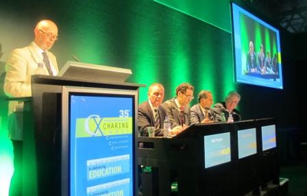
At today’s CX Renal Denervation Session Great Debate, delegates voted 3:1 against the motion that renal denervation should be used routinely to treat hypertension. As all of the speakers, regardless of which side of the debate they were, agreed that renal denervation should only be used to treat patients with resistant hypertension, the real focus of the debate centred on what was meant by the word “routine”.
Mel Lobo (London, UK), who was supporting the motion that renal denervation should be part of routine clinical practice to treat hypertension, claimed that the definition of routine was a “key issue” in the debate. He explained that patients who were suitable candidates for the intervention belonged to a highly selective group of patients, who had true resistant hypertension (ie. non-concordance with medication and other causes of resistant hypertension had been ruled out) and who would be managed by hypertension specialists rather than by primary care physicians. Lobo added that, in his view, renal denervation should become “step five” of the UK National Institute for Health and Clinical Excellence (NICE) pathway for the management of hypertension, commenting: “We do not have any randomised controlled trial data for step four [the use of spironolactone; only observational data are available], so why not have renal denervation as step five? Believe me when I say adding in a sixth or seventh antihypertensive drug is not going to work.”
Felix Mahfoud (Homburg, Germany), who was Lobo’s co-proponent of the motion, agreed that the routine use of renal denervation referred to it being used only in the “very specialised” group of patients with true resistant hypertension. He said: “Is it really ethical not to offer patients a modality that could benefit them?”
Speaking against the motion, Mark Caulfield (London, UK) disputed that “routine” could refer to an intervention being used in a highly selected group of patients. Caulfield commented: “Routine use means everybody; it implies that the intervention should probably be used in a large number of patients. Renal denervation is not ready to be used in routine clinical practice because there is not evidence base to support it being used in this way.” He added that the NICE consensus document on renal denervation, of which he was a co-author, was specifically drawn up because there were concerns that the doctors would use the intervention for all patients with hypertension rather than just those with true resistant hypertension.
Also speaking against the motion was Bryan Williams (London, UK), who said he was doubtful about the data from renal denervation studies. He commented: “What is critical is how many patients in these studies were actually taking their antihypertensive medication? If the drop in blood pressure observed in these studies is really a result of renal denervation on top of antihypertensive medication, then that is impressive. But if it is actually a result of patients starting to take their drugs [because they are now in a clinical trial], then it is not impressive.”
Lobo responded by saying there had to be element of trust and that “We have to believe our patients when they say they take their medication. We are not able to follow them home and check up on them.”
At the end of the debate, 3:1 delegates voted to support Caulfield and Williams that renal denervation should not be part of routine clinical practice for managing hypertension. Summing up the data, the chair of the session Neil Poulter (London, UK) said he hoped that by the time of CX36 (12–15 April 2014), more data on renal denervation would be available and the issue could be further discussed.
Endovascular management of ascending aortic pathology
By Ralf Kolvenbach
The incidence of thoracic aortic aneurysms is estimated to be as high as six cases per 100,000 person-years, and replacement of the ascending aorta accounts for the majority of cardiothoracic aortic procedures.
Aneurysms and dissections of the ascending aorta are still mainly treated operatively with cardiopulmonary bypass. Ascending aortic aneurysms with normal sinuses and aortic annulus require only replacement of the ascending aorta from the sinotubular ridge to the origin of the innominate artery with a Dacron tube graft.
Due to the significant morbidity and mortality of these procedures high-risk patients are not considered for cardiac surgery. There are a number of anecdotal reports describing endovascular stent grafting of various pathologies including deployment of a fenestrated stent graft in a case of an ascending aortic rupture.
Endovascular treatment of the ascending aorta is particularly challenging because of the anatomical features of this aortic segment. Acute and chronic Type A dissections can be treated with a tubular endograft when the aortic valve and the coronary arteries are not involved. An analysis in our clinic of 30 consecutive patients admitted with ascending aortic pathology showed that in 75% ascending aneurysms had a conical shape without a proximal landing zone. Only in a few cases the ascending aorta had a tubular configuration with a long proximal landing zone of at least 2cm. This makes over-sizing and an optimal graft configuration particularly important. Only patients without connective tissue disorders, clinically relevant aortic incompetence, stenosis or concomitant coronary artery disease can be considered for an endovascular procedure.
Material and methods
So far only patients with ascending aortic pathology who were considered unfit for open surgery were treated with an endograft. We also excluded patients with malperfusion or any preoperative unstable clinical condition after Type A dissection. Also excluded were patients with severe aortic valvular disease including aortic valvular incompetence as a consequence of an acute Type A dissection, coronary artery disease requiring surgery, and any kind of connective tissue disorder. Included in our ongoing study are patients with intramural hematoma, floating thrombus after chronic Type A dissection and penetrating aortic ulcers. Also included are aneurysm patients without aortic valve incompetence or significant dilatation of the aortic annulus requiring composite graft replacement of the aortic valve and the ascending aorta.
Patients with asymptomatic penetrating aortic ulcers (PAU) were initially managed conservatively. If symptoms did not resolve, or patients continued complaining about chest pain, endovascular exclusion was discussed with the patient.
Threshold for aneurysm patients is an aortic diameter of 6cm or larger and an adequate landing zone proximal to the coronary arteries. Patients were excluded if they were good candidates for open surgery. In cases without a good landing zone an aortic banding procedure using a mini sternotomy was performed prior to stent graft deployment as originally described by the Zurich group of Lachat.
The length of the stent graft was depending on the length of the outer curve of the ascending aorta which was regularly longer than the distance determined by center line measurement.
All operations were performed under general anesthesia. In addition to our monitoring protocol for TEVAR transoesophageal ultrasound was performed to control cardiac and valvular function. Cardiac arrest was induced with adenosine administration when required. A temporary ventricular pacemaker was placed through the jugular vein.
After road map angiography selective coronary angiography was performed to outline the origin of the coronary arteries.
An ultra stiff guide wire (Lunderquist, Cook) was placed into the left ventricle after passage of the aortic valve with a vertebral catheter over a 0.035 guide wire. The endograft (Cook custom made ascending endograft) was carefully placed across the aortic valve into the left ventricle. The kind of graft used depended on the size required.
Tracking of the stiff wire all the time during the procedure was essential to avoid ventricular perforation. At the end of the procedure ventriculography was performed to rule out any damage to the left ventricle or the valve apparatus. In case of any ECG changes coronary angiography was added before removing the catheter and sheaths. Before discharge a contrast enhanced angio CT was performed as well as cardiac ultrasound examination.
Ascending aortic dilatation may be caused by intrinsic pathology of the aortic wall, or hemodynamic factors caused by a stenotic aortic valve: High velocity and turbulent flow downstream of the stenosis place mechanical stress on the aortic wall.
The thin wall of the ascending aorta does not permit aggressive over dilatation. The graft was deployed in most cases without any additional balloon dilatation. The walls of the aortic sinus (sinus of Valsalva) are considerably thinner than the wall of the aorta. Hooks or bear springs should therefore be deployed in a safe distance from the sinus and the origin of the coronary arteries to avoid erosion and perforation. One of our first patients suffered from a stroke postoperatively.
Calcifications and thrombus lining of the ascending aorta and aortic arch were the main parameters for increased stroke risk
Access can be a problem since in most aneurysm cases a 24F access sheath was required. In two cases with sever calcification of the iliac vessels we had to use the left carotid artery as an access which proved to be uneventful. Alternatively using a small thoracotomy the endograft can be deployed through the apex of the heart.
There is so far only one dedicated graft for the ascending aorta (Cook). Since most aneurysms have conical shape a stent graft designed for Type a dissection cases is not necessarily suitable for aneurysm patients. So far the anchoring zone of a few millimeters only, requires fixation with hooks, significant over sizing and coil deployment into the sac of the aneurysm if necessary. Banding of the ascending aorta is an option to increase the landing zone in aneurysm patients with conical aneurysm morphology.
The aortic impulse which is directly proportional to the mean blood pressure, the cross sectional area of the aorta and the duration during systole varies inversely with the distance from the aortic valve. In stent grafting of the aortic arch and the descending aorta high aortic impulses can cause significant pulsate motion of the arch and inadvertent movement of the stent graft. In the future more active fixation with a stapler would probably permit safer deployment, less Type I leaks and better long term performance. Especially when treating more advanced stages of aortic pathology a valve bearing conduit with fenestrations for the coronary arteries will be necessary.
Before a routine use can be advocated outside of studies several issues must be addressed. There should be dedicated grafts for ascending aneurysms and for acute dissections like the one available so far. Precise deployment of the graft adjacent to the coronary arteries is essential in aneurysm patients. In most cases there is a landing zone of a few millimeters only, deployment too distal from the sinotubular junction results in kinking of the graft and subsequently a type I leak.
We can conclude that stent grafting of the ascending aorta is technically feasible but should be reserved to selected high risk patients only, preferably in centers where vascular specialists cooperate closely with interventional cardiologists. Cardiac surgery with cardiopulmonary bypass and if necessary deep hypothermia is still the gold standard when treating ascending aortic aneurysms though still associated with significant morbidity and mortality. Stent graft exclusion of more advanced and complex ascending aortic aneurysms should be reserved for high risk patients only in centers with the necessary skills to perform transvalvular cardiac procedures. This may change in the future when more dedicated grafts become available. The future will and has to show valve bearing endovascular conduits which will permit minimal invasive treatment of most aortic aneurysms. The grafts available so far already permit emergency endovascular treatment of type A dissections. Yet in aneurysm patients compromises are still necessary.
Clinical need drives intraoperative imaging to the next level

For the very first time, three full-scale sterile hybrid operating suites from GE Healthcare, Philips and Siemens were on display at CX. The importance of quality imaging as a prerequisite for improved clinical outcomes was emphasised in every section of the main programme. This went hand in hand with calls from physicians for high-quality image availability in the intraoperative setting.
Imaging is certainly at the heart of endovascular intervention, and it is now widely accepted that using the best available imaging can have a direct impact on achieving the best clinical results. With the blurring of boundaries between specialties in the endovascular arena, there was a clear need expressed by clinicians at CX for improved imaging at the intraoperative stage. Delegates at CX35 are split in nearly equal proportions along the disciplines of vascular surgery, interventional radiology and interventional cardiology.
Georg Nollert, director, Global Marketing, Siemens, told CX Daily News: “I am very happy that the focus on imaging is increasing. In the past, vascular surgeons were satisfied with inferior image quality and other interventionalists such as radiologists and cardiologists benefited from using the best available imaging. I believe that surgeons ought to have the same image quality in order to get the best results.”
In the preoperative and postoperative setting, ultrasound and other sophisticated imaging modalities such as CT or MRI are widely available. Nollert said: “Intraoperatively, however, imaging was limited to the use of C-arms (2D fluoroscopy) in the past. Therefore, one available solution was to enable the superimposition of the preoperative images with the intraoperative images. Using preoperative CT or MRI, this then creates the 3D road map that interventionalists could use for very sophisticated interventions.
“Another possibility was just to use intraoperative cone beam CT, and we are getting close to conventional CT quality with this. The elegance of this solution is that the images are automatically registered to the patient and there is, on the other hand, the actual anatomy of the patient on the table that is probably not the anatomy that you have on the [preoperative] CT (because he is in a different position and may also be in a different state of hydration). We therefore believe that the most precise imaging is done intraoperatively, particularly when you have inserted wires or other devices that have changed the anatomy. This might bring up the issue of distortion/deformation of the vessels which can be quite substantial and the overlay could somehow suggest that the operator is in the right lumen, even if they are not.”
Nollert told CX Daily News that in the past, the issue with cone beam CT was one of image quality, particularly with reference to contrast resolution and that Siemens has been working on improving this aspect. “We now have new algorithms to reduce metal artifacts and also better detectors in the latest family of systems, the Artis Q. Artis Zeego also has the latest technology and the latest detectors and we have increased the contrast resolution for cone beam CT substantially. It is important to bear in mind that all cone beam CT is not equal. We have new protocols that result in the quality being much superior to regular/conventional CT. However, image quality for cone beam CT depends on reconstruction algorithms, and how you deal with artifacts and distortion, and those algorithms are widely different in the market.”
He highlighted that the Artis Zeego was a flexible, robotic system that can adjust to the table and to being used by the whole team in the operating room. The system also allows for maintenance of the sterility and environment of the operating room as it keeps the ceiling free for uninterrupted laminar flow, or use of operating room lamps, for example. “With regard to 3D capability, the Artis Zeego has some absolutely unique features and can image large volumes, fast and achieve a superior image quality,” he said.
Clinicians in the session made the point that the robotic arm with the automated motion had the drawback that the user could not always predict how the system was going to move and often found that it was quite hard to anticipate the movement.
Kirsten Zuurmond, clinical scientist, Philips Healthcare and Koen Noordermeer, Business Development Manager, Philips Healthcare, told CX Daily News: “Philips is very concerned about the quality of intraoperative imaging. In order to reduce radiation dose and contrast values, Philips offers the possibility of fusion imaging where clinicians use the preoperative CT as a navigational map during the procedure. We also offer the flexibility of working with cone beam CT, such as in emergency situations, so that it is possible to make an accurate 3D image at the intraoperative stage.
They highlighted the flexibility of positioning the c-arm Philips system within the hybrid operating room, the advantages offered by the new system Alluraclarity, in achieving significant dose reduction, without compromising on the image quality available to physicians.
3D superimposition on fluoroscopy is not yet ideal
During the Monday morning session the issue of distortion resulting from overlaying preoperative 3D images intraoperatively was discussed. A panel comprising of chairman Peter Taylor Tara Mastracci, Cleveland, USA, Krassi Ivancev and Ian Loftus, both London, UK, noted that with fusion imaging, there was no compensation for vascular distortion and that there was imprecise fusion usage for endograft positioning and cannulation of target arteries.
“One of the issues that many clinicians have with fusion imaging is its lack of responsiveness to deformation, but most believe that a fix is on the horizon,” Mastracci said. Sixty two per cent of the audience in the session also voted against the motion that fusion of preoperative datasets is ideal.
Zuurmond highlighted that clinical opinion was clearly divided on the topic with some clinicians experiencing a high degree of accuracy. She then also explained that the company was focusing on the ostia as a target in fenestrated EVAR procedures and noted that they hoped to provide the extra help that could be useful in the area.
Noordermeer then added: “We see the benefit of the current technology but are of course working on improvements for the future to make it even more accurate and applicable. We are interested in is bringing solutions that are easier to use in the hybrid operating room and are working on automating workflow steps, making access easier and providing stepwise guidance for physicians.”
Ease of use
Gregory McIff, global director, Cardiovascular Strategic Marketing and Delphine Germain, global product manager for Hybrid OR, GE Healthcare, told CX Daily News that flexibility in the operating room, ease of use for physicians and advanced image quality were optimised to the best degree that technology allowed today in the GE systems.
In response a questions about image quality not being as good as it could be at the time of intervention, McIff noted that image quality was subjective to the user. “We know from technology that a flat panel detector gives a clearer image than an image intensifier system, so you are going to see some changes in that. But when you are dealing with aortic interventions the ability of a system to provide good imaging is perhaps, adequate. You can always have better imaging—I would love to drive a Rolls Royce, but I have to make do with my Chevrolet!”
Germain said, “We definitely emphasise that the system is easy to use. You can have the best thing in life, but it has got to be accessible, not complicated to use, too. This is an area we will keep investing in: improving the ease of use. We already have systems that allow surgeons to learn fusion imaging from tableside. With regard to superimposing preoperative imaging data within the Hybrid OR, the technique is getting complicated and it is important for us to make sure that people can use it on a routine basis.”
Both highlighted that GE focused on combining the best of the hybrid operating room with the best of the advanced imaging techniques. They drew attention to the fact that the Discovery IGS 730 was not mounted either on the wall or on the ceiling but that it could move freely in the room.
“The system confers the benefit of mobile systems that can move in the room freely so that clinicians can have access to patients, and at the same time have the ability to be brought back into imaging position and have all the advantages of a fixed system in terms of imaging such as 3D imaging and 3D fusion,” they said.
CX ilegx Collaboration embraces Electronic Endovascular Education
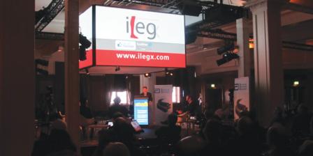
This year, the ilegx multidisciplinary team, continuing in its aim to reduce the number of leg amputations, broadcast—for the first time—edited live below-the-knee endovascular procedures to the Far East, in association with Abbott Vascular. Also, as part of the ilegx programme, the King’s College Hospital Open Access System showed the latest treatment options for diabetic foot. Innovative methods of foot revascularisation were also discussed and a guidewire tutorial was taught.
The morning session titled “Electronic Endovascular Education—Edited live cases broadcast online to the Far East”, chaired by Max Amor (Essey-les-Nancy, France) and Roger Greenhalgh (London, UK) showed delegates three complex below-the-knee procedures from Germany, Italy and France.
The first case was performed by Andrej Schmidt in December 2012 in Leipzig, Germany. The second procedure was carried out by Roberto Ferraresi in March 2013 in Milan, Italy, and Eric Ducasse undertook the third case in Bordeaux, France, in March 2013. Ducasse said: “In the past decades we have seen major advances in the treatment of below-the-knee lesions with dedicated materials and retrograde approaches to peripheral arterial disease.” For this case, Ducasse showed below-the-knee techniques using Abbott Vascular materials. He showed a retrograde approach through the peroneal artery followed by guide wire proximal recapture and successful balloon angioplasty.
After each case, Dierk Scheinert, Leipzig, Germany; Flavio Airoldi, Sesto San Giovanni, Italy; Ferraresi, and Ducasse, answered questions, via video link, from delegates watching in the Far East. Questions came from Japan and India, and also from Tunisia.
At the end of the session, Greenhalgh told delegates: “This Electronic Endovascular Education session has been a wonderful experience with great educational value. I have to thank these physicians from Germany, France and Italy for those fantastic results and for sharing with us all that can be done to save legs.”
He added: “This experience forms part of the CX ilegx Collaboration Day and ilegx stands for interdisciplinary management of legs because we are concerned that too many legs are being amputated,”
Greenhalgh told CX Daily News: “In these times of economic difficulties—when flight costs are on the rise—this online educational experience could be a cost-effective way of sharing education. We would like to invite the participants of this experience to share their opinion via twitter or facebook on whether this should be a pattern to be followed in future CX meetings”
King’s College Hospital Open Access System aims to save diabetic foot
The King’s College Hospital Open Access System, London, UK, “includes a multidisciplinary team of podiatrists, nurses, microbiologists, vascular surgeons, orthopaedic surgeons, diabetologists and interventionalists dedicated to providing an urgent, immediate treatment to patients who are at risk of developing necrosis, gangrene and losing their legs,” Michael Edmonds, King’s College Hospital, told CX Daily News. “The two drivers for this—in diabetic patients—are infection and ischaemia. Rapid diagnosis of infection and rapid treatment will prevent the progression of the necrosis. At the same time, the vascular system should be addressed and we should go forward with revascularisation, either with angioplasty or bypass, depending on the degree of the circulation problem as soon as possible,” he added. Edmonds presented an update entitled “Rapid referral and treatment within the concept of the diabetic foot attack” at the CX ilegx session.
Physicians with different specialties from the King’s College Hospital Open Access System also gave presentations at the ilegx Collaboration Day on their experience treating diabetic foot as a multidisciplinary team.
Jason Wilkins, London, UK, presented a modern interventional approach to the diabetic foot. He told delegates, “The modern interventional approach to the diabetic foot begins with teamwork and recognition of patient-centred care being at the forefront of the team approach. Ischaemia with neuropathy or infection is considered an emergency and robust patient pathways are mandatory in providing timely intervention.”
Wilkins highlighted that revascularisation was a basic requirement for successful treatment and amputation prevention. “Revascularisation may be surgical, radiological or a combined approach according to the presentation and nature of disease and distribution,” he commented.
According to Wilkins, modern techniques and equipment provide the interventionalist with excellent tools for revascularisation with angioplasty, stenting and recanalisation of multiple long occlusions. He said: “The understanding and availability of modern equipment and techniques along with an effective multidisciplinary and timely approach to urgent revascularisation result in improved outcomes for our patients.”
Hisham Rashid, vascular surgeon, London, UK, presented “Distal and ultra-distal bypass: a discussion on the foot angiosomes—fact or fiction?”
Rashid told delegates that the angiosome concept was developed in 2006 by Attinger. He commented on a study—in press—that he undertook to evaluate the impact of the angiosome concept in a group of 142 diabetic and non-diabetic patients who underwent distal and ultra-distal bypass surgery for critical limb ischaemia with significant foot tissue loss. Rashid reported: “In this cohort of patients the healing and time to healing was not affected by the angiosome revascularised, but was significantly affected by the quality of the arterial pedal arch. In patients with no pedal arch, the healing was significantly slower and inadequate compared to the complete and incomplete pedal arch subgroups. However the amputation-free survival rates were similar in all groups.”
International input to limb salvage
Also, as part of the Kings College Hospital Access System programme, speakers from Europe and USA gave their views and experiences on limb salvage.
Carlo Setacci, Siena, Italy, talked on the latest guidelines on diabetic foot treatment: The Italian consensus document. Carlo Caravaggi, Milan, Italy, presented a new integrated surgical approach—based on timing—to reconstruct the diabetic foot. Christopher Attinger, Washington, USA, told delegates about surgical care of the wound with debridement and planning of amputations and reconstruction. An interventional approach to the diabetic critical limb ischaemia patients vs. non-diabetics was presented by Roberto Ferraresi, Milan, Italy. David Armstrong, Tuscan, USA, spoke on techniques to correct foot deformity by surgical means.
Revascularisation challenges
In the afternoon, Roger Greenhalgh, London, UK, chaired the session on revascularisation challenges on the treatment of critical limb ischaemia.
Frank Vermassen, Ghent, Belgium, highlighted the importance of keeping vessel patency in the long run. He said: “Sustained patency of the wound-related artery is mandatory to optimise the chance for wound healing, to avoid repeat intervention and to preserve the limb.” Thomas Zeller, Bad Krozingen, Germany, said: “Patency is necessary but not sufficient for wound healing and ultimate limb salvage.” He added, “Drug-eluting balloons may be the solution to achieve the necessary patency levels within the extensive multivessel arterial disease typical of critical limb ischaemia.”
In the discussion, the question of using more than one balloon to achieve the necessary patency in critical limb ischaemia patients was raised. However, this approach would increase costs. Greenhalgh made the point that only patients with insurance companies willing to pay for this and patients who can afford it would receive the treatment.
A completely percutaneous closure approach is feasible in most cases
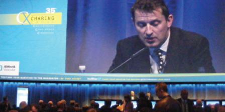
At the Abbott Satellite Symposium, yesterday, Ian Loftus (London, UK) reviewed the data for percutaneous closure in endovascular aortic procedures, stating that the percutaneous closure approach was “technically feasible in most cases, is safe, and leads to early mobilisation and reduced length of stay”
Loftus explained that with endovascular aortic aneurysm repair (EVAR), the traditional femoral cutdown for vessel access was still used in many cases. He commented: “Studies have demonstrated groin complication rates ranging from 5% to 15% associated with femoral cutdowns, including wound infections and lymphatic leaks. This can hinder recovery and lead to prolonged length of stay.”
He added that a percutaneous approach using arterial closure devices could potentially avoid many of these complications. Loftus reported: “Previous series and a single small randomised trial have demonstrated high technical success rates and significantly shorter operation times, quicker time to ambulation, reduced length of stay and reduced overall procedural costs associated with using the Abbott Prostar XL device [for percutaneous closure].”
According to Loftus, data from a US multicentre randomised clinical trial comparing percutaneous closure using the Proglide closure device (Abbott Vascular) with standard surgical cutdown found that major access site complications were significantly lower in the percutaneous group (6% vs. 10% for surgical cutdown) and that minor complications were halved (4% vs. 8%). He added: “There was also a significantly shorter procedural time (106 vs. 141 minutes) and time to haemostasis (10 vs. 23 minutes).”
Concluding, Loftus stated: “Complete percutaneous treatment is technically feasible in most cases, is safe and leads to early mobilisation and reduced length of stay”. He commented that the complication profile of this approach was different form the open approach, but said complications could be minimised with “prudent case selection, training and careful device deployment.”
Also at the “Close with confidence” symposium, Matt Thompson (London, UK) spoke about “tips and tricks” for percutaneous closure. These tips included being aware of blood clots, the importance of being patient, and advice on accessing the common femoral artery. He said: “Getting the correct puncture site is absolutely key to a successful procedure. Ultrasound is the best way of ensuring accuracy of puncture.” He concluded that the percutaneous approach was now the default approach for all endovascular procedures at his centre (St George’s Vascular Institute, London) and they had seen an improvement in patient outcomes after adopting the approach. Thompson added there was “definitely a learning curve” with the percutaneous closure approach.