


CX NEWS
Spotlight on Bolton Medical’s Thoracic Arch Branch device
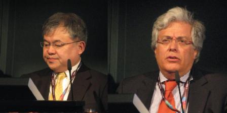
In a Bolton Medical sponsored event, Piergiorgio Cao, Rome, Italy, and Toru Kuratani, Osaka, Japan, both presented their experience with the company’s Thoracic Arch Branched Device. Kuratani also presented his experience at the CX Complex Edited Live Cases and Case Reports.
Cao spoke to delegates about complex cases in which deployment of the Thoracic Arch Branch device was successful using single- and double-branches. Cao showed 3D CT images that indicated the successful use of the device in single branches in five cases (including ascending and descending aorta dilatation, arch aneurysm, pseudoaneurym, and endoleak amongst other comorbidities such as diabetes and hypertension). He commented that final angiograms of these cases were good.
With the double-branch device, Cao spoke about one patient with a thoracic aortic aneurysm involving the left subclavian artery with no proximal neck posterior to the left circumflex coronary artery (the left circumflex coronary artery and the iliac artery were also very close together). He reported that this was successful but the patient had a type 2 endoleak, which was due to be treated in the coming weeks.
Referring his experience with the device, Kuratani reviewed 3,015 cases of aortic aneurysms. Of these, 2,383 had been treated with a stent graft. He also showed a video to delegates of his team performing a debranching thoracic endovascular aneurysm repair with the Bolton Medical device.
The arch branch technology (based on the Relay non-bare stent plus platform, intended for zone 0 deployments), according to Bolton Medical, incorporates a proximal clasping mechanism intended for repositioning and a V-patch inner sheath for expansion and optimal alignment.
The (single and double) branch graft is expected to be examined via data collected by the company, from further clinical experience and a trial of the branched devices from centres preforming thoracic abdominal aneurysm repair. In phase I of this analysis, the single branch device will be tested in five institutions in six cases. In phase II, the double branch device will be assessed in six cases across six institutions.
New three-stage hybrid graft reviewed
Heinz Günther Jakob (Essen, Germany) told CX delegates about a new three-zone aortic arch hybrid prosthesis, which has been designed to reduce ischaemic times by 50%. Jakob reported that the three-stage hybrid graft was created with the aim of reducing the rate of complications seen with the E-vita open hybrid prosthesis (Jotec) in some patients.
He said that it consists of a “descending aortic covered stent graft, followed by a non-covered stent graft for the arch, and a Dacron part for the ascending aorta” and that it has been designed to “reduce ischaemic times by more than 50%.”
The primary indications for the graft would be “sick octogenarians” or in situations where there is “dramatic malperfusion” (such as myocardial infarction, stroke, and prolonged visceral ischaemia). He reported that in a small pig study (of six pigs), there was a technical success rate of 100% and a visceral ischaemia time of 21 minutes.
Jakob concluded: “After passing all regulatory affairs with the hospital, four grafts are under construction for the first-in-man implantation.” He added at present, the graft was just designed to be implanted via conventional surgery (through median sternotomy), but said that “of course” they also planned to assess the transapical or transfemoral route as well.
LIVE from CX35: Restore II registry confirms safety and efficacy of Relay devices
Today, Martin Funovics (Vienna, Australia) presented data from the Restore (Relay endovascular registry of thoracic diseases) II registry, which he said confirmed the safety of the Relay and Relay NBS thoracic stent grafts (Bolton Medical) in thoracic endovascular aneurysm repair (TEVAR)
Funovics explained that Restore II was a prospective worldwide registry of 234 patients who have undergone TEVAR with a Relay device. He reported that the Relay device was available as a “clasped” version with conventional bare springs and as an unclasped version with covered springs (Relay NBS). Referring to the unclasped version, Funovics said: “This version for the first time has differential attachment modalities for the inner and outer curvature.” The inner two clasps, he commented, that are guided by long levers from behind rather than proximally. “During the opening process, these two levers can slowly and in a very controlled manner guide the inner curvature apeses to the aortic wall.” He added that the device has been specifically designed for the descending aorta.
The patients in the registry, Funovics reported, reflected the “typical TEVAR patient population”, but he added they did have a “substantial amount” of cardiovascular comorbidities compared with the typical TEVAR population.
Funovics commented that with the Relay and Relay NBS devices, the technical success rate was 95.3% and there was a very low rate of systemic complications. Additionally, the rate of mortality following the procedure was similar to other series.
He concluded: “The Restore II registry confirms the safety and efficacy of our differential detachment mode of the inner and outer curvature springs. The results were comparable, especially in terms of neurological complications, to other registries and a relative high rate of technical success was achieved.”
LIVE from CX35: STABLE shows favourable two-year results with Zenith dissection system in type B dissections
Joseph Lombardi, Philadelphia, USA, presented two-year results of STABLE (The study of thoracic aortic type b dissection using endoluminal repair) today. STABLE, he said, is a prospective, non-randomised, multicentre clinical study conducted at investigational sites in Europe, Australia, and the United States to demonstrate the safety and effectiveness of the Cook Zenith Dissection Endovascular System in the treatment of patients with type B aortic dissection.
Developed as a less invasive alternative to open surgical repair and specifically for the treatment of type B aortic dissections, this system comprises the Zenith TX2 Thoracic Aortic Aneurysm Endovascular Graft with Pro-Form and the Zenith Dissection Endovascular Stent. “The combined use of proximal stent-graft and distal bare stent components provides a means to seal the primary entry tear and support the necessary length of dissection without risk from coverage of branch vessels,” Lombardi said.
A total of 86 patients (73% male, mean age 59 years) were enrolled in the STABLE study. More than half of the patients (64%) were treated in the acute phase (within 14 days of symptom onset) and a majority (73%) had presenting symptoms of impending aortic rupture and/or branch vessel malperfusion.
Lombardi, global principal investigator of the study, presented study results through 24 months, reflective of data received as of March 2013. The overall 30-day mortality rate was 4.7% (4/86), and Kaplan-Meier estimates of patient survival were 88% at 12 months and 85% at 24 months. During follow-up through two years, five patients experienced aortic rupture and no patient required conversion to open repair. There were seven cases of stroke (six cases within 30 days) and one case of paraplegia (within 30 days). Renal failure occurred in nine patients (none required permanent dialysis) and retrograde dissection occurred in seven patients (two patients died; four underwent re-interventions). Aortic remodeling, indicated by an increase in the true lumen size and a decrease in the false lumen size, was observed in both the descending thoracic aorta and the distal abdominal aorta.
“These results continue to indicate favourable clinical and anatomic outcomes with the use of a composite TEVAR construct. Follow-up through five years is ongoing to assess long-term effectiveness of this treatment strategy,” Lombardi stated.
CX35 experts sharply divided on type II endoleak challenge
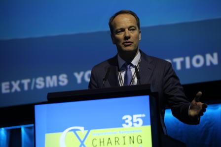
The management of type II endoleak provoked a whole host of opinions among experts at CX35 yesterday. Are type II endoleaks benign, or not? Are “dangerous” type II endoleaks really misdiagnosed type I or type III endoleaks? Do type II endoleaks need treatment (and how), or is leaving them akin to “leaving a baby on a railway line”? Experts taking part in a panel discussion about the management of type II endoleaks following endovascular aneurysm repair (EVAR) did not agree
According to Hence Verhagen (Rotterdam, The Netherlands), type II endoleaks by themselves are benign and there is no proof that type II endoleaks cause type I or type III endoleaks. He said: “With a type II endoleak, it may be the outflow vessel that you are looking at instead of the inflow vessel [ie, it is the result of a type I or type III endoleak].”
Verhagen claimed that there “was no need to worry” about the treatment of type II endoleaks because they were associated with low pressure. He added that even if they were treated, there was little evidence that treatment would be effective—“In 100 patients with a type II endoleak, only two to five have a growing sac. Of these, only 30% will be successfully treated and that is the rate reported with experienced centres; therefore, only one of 100 patients will benefit from treatment.
However, Jean-Pierre Becquemin (Créteil, France) disagreed and commented: “I am really convinced that sometimes if you wait for more than five years to intervene, that type II endoleaks will lead to sac enlargement and maybe a type 1 endoleak.”
Becquemin argued that certain patterns of type II endoleaks were not benign and “must be treated by all appropriate means before catastrophe occurs”. He explained that he and his colleagues, in a recent study published in Journal of Vascular Surgery, reviewed the long-term outcomes of consecutive patients who had undergone EVAR for atherosclerotic infrarenal aortic or aortoiliac aneurysms between June 1995 and May 2010 at their centre (Henri Mondor Hospital, Creteil, France).
After a mean follow-up period of 31.3 months (range 12.4–61.4 months), 201 patients (of 700 overall) had at least one type II endoleak and these patients were at higher risk of sac growth and re-intervention compared with patients without a type II endoleak. Additionally, persistent (p<0.001) and recurrent (p=0.008) type II endoleaks were both highly predictive of sac growth as were type II endoleaks that were associated with a type I or type III endoleak (p<0.001). Becquemin reported that mortality was not increased in patients with type II endoleaks but added: “Type II endoleaks did not kill these patients because these endoleaks were treated.” Concluding the results of study, Becquemin said: “Believing type II endoleaks are benign is like believing leaving a baby on a railway line is fine”—ie, there is no immediate danger, but danger may be approaching.
In the ensuing discussion, Becquemin acknowledged that type II endoleak were not necessarily the problem per se, and that it might actually be sac growth that was the main issue. “But you cannot neglect type II endoleaks. That is my strong feeling,” he said.
Matt Thompson, London, UK, claimed that there was not a “one size fits all” answer regarding whether or not type II endoleaks were benign. He said: “I am a pretty firm believer that some type II endoleaks are dangerous and that they are going to lead to aneurysm problems with the endograft, but I think the vast majority are benign.” He added that, probably, the “biggest problem” with type II endoleaks was that some endoleaks were labelled as type II endoleaks when “in reality”, they were actually a high-pressure type I or type III endoleak. “I think that is where the confusion comes from”, he commented. In his view, multi-imaging modality was “very important” to prove that sac growth was really being caused by a type II endoleak rather than a type I or type III endoleak.
Thompson also claimed that his experience with open aneurysm repair indicated that some type II endoleaks could lead to sac expansion. CX35 programme chairman Roger Greenhalgh (Imperial College, London, UK), who was chairing the session, commented that he had also seen open surgery cases that had showed type II endoleaks to be the cause of sac expansion.
He said: “Years after a perfectly good Dacron replacement by my predecessor, an apparent second rupture occurred. When I got it controlled and opened the sac, the whole thing was being driven by what today we would call a type II endoleak, with a massive lumbar, which was forcing the sac and I had to tie it.” He added that while not all type II endoleaks would lead to rupture, he was concerned about the possibility of type II endoleaks driving sac expansion.
In his presentation, Thompson also gave an update about the Nellix technology (Endologix) for EVAR. He said that, at present, there was not much long-term data for the technology but the data so far did indicate it could represent a “paradigm shift” in treatment because of its apparent ability to reduce complications and endoleaks.
Although the panel discussion did not reach a consensus, there was a clear message from the audience—nearly 80% of delegates voted against the motion that “Type II endoleaks without sac expansion of more than 5mm per year needs intervention”. Greenhalgh commented: “By implication, some of you believe that type II endoleaks do need intervention if there is sac expansion greater than 5mm.”
Apples and oranges
Robert Morgan (London, UK) said that type II endoleaks were “heterogeneous” and that comparing the different types of type II endoleak was like “comparing apples and oranges. Some type II endoleaks are ‘more benign’ than others.” He added that in type II endoleaks, absence of mural thrombus, the size of the endoleak nidus, the presence of inflow and outflow vessels, and the size of lumbar arteries or the inferior mesenteric artery were all factors in predicting future sac enlargement.
However, he claimed that studies had indicated that type II endoleaks were associated with a low rate of rupture.
Morgan also reviewed the treatment of type II endoleaks, stating that there was not enough data to determine the direct sac puncture embolisation technique was the preferred option to the transarterial technique approach. He said: “In practice, there are enthusiasts for either technique. Logistics [in my view] favour a transarterial technique.”
According to Paolo Frigatti (Udine, Italy), preventing—rather than treating—type II endoleaks might be a valid strategy. He said that previous reports had shown: “Injection of fibrin glue alone or in association with microcoils in the aneurysm sac during EVAR can facilitate sac thrombosis and reduce the incidence of type II leaks during follow-up.”
Imaging for type II endoleaks needs improvement
Frans Moll (Utrecht, The Netherlands) said that imaging needed to be improved to detect more type II endoleaks. Improved imaging would also detect more feeding vessels for better embolisation. He said that MRI with blood pool contrast agent might give the needed improvement. However, he added: “Not all patients and stent grafts are MRI compatible and nothing of the effect of this better imaging on the outcome of EVAR is proven yet.”
Taking into account the diverging views on the management of type II endoleak, Greenhalgh said: “I wish I could say to the audience that the speakers today have made it easier, but perhaps it has been true to say, it is has been made more difficult for you. But one thing always to remember is that if you do not know the answer, clock that you don’t know the answer—so that you try to harder to find an answer.”
CX35 type II endoleaks survey
Delegates were invited to be part in a CX35 survey about the management of type II endoleaks yesterday. Please visit the BIBA MedTech Insights stand (Gallery Level) if you would like to participate.
Latest technology presented at the CX Innovation Showcase
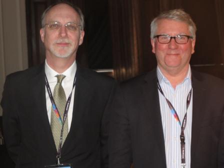
Nick Cheshire and Stephen Greenhalgh, London, UK, chaired the CX Innovation Showcase today. The audience heard about innovation challenges and initial results from a series of new technologies for abdominal, thoracic, lower limb, neurovascular and venous interventions. Results for the new Crux vena cava filter were also presented.
Greenhalgh spoke to the audience about innovations and challenges to innovation in collecting data. He presented insights from the European Vascular and Endovascular Monitor, a consumption-based monitor covering 200 centres from Western Europe. He said that the advantage of a monitor approach was that it was enduring and effective in rapidly-shifting markets.
From the thoracic market, data showed that thoracic endovascular aneurysm repair remains the “gold standard” and that the current issues in the thoracic field were the size of the aneurysm, and the perceived need for a screening programme. For carotid procedures, Greenhalgh said the data indicated open surgery as the gold standard and that endovascular procedures were focused in the German and Italian markets.
“Endovascular procedures continue to grow at the expense of open surgery,” Greenhalgh commented on aortic-iliac procedures and femoropopliteal, with the use of drug-eluting balloons on the increase in the latter. Hefsyv identified the key trends in the market and stated that, “The biggest challenge is doing this in a cost effective way”.
In a following presentation, Caroline Hough, London, UK, spoke about usage and attitudes as customised studies. She said that customised research provides answers to specific questions such as the effect of a new market entrant, understanding purchasing decisions, factors impacting competitiveness of a product vs. the competition. She added that the research is tailored to the client and can be based on specific demographics and on qualitative and quantative research. She also noted that qualitative data work by “drawing out themes”.
She presented a case example of how targeted quantitative analyses addressed the type II endoleak challenge at CX35. The aim was to understand the current level of knowledge of type II endoleaks and to assess prevalence of treatment and preferred treatment protocols. The methodology was to target a global audience of physicians attending CX35 face-to-face and with a hard-copy quantitative questionnaire.
The results will be collated and edited highlights will be published in Vascular News and Interventional News.
Crux vena vena cava filter
Among the devices that were featured at the CX Innovation Showcase, Andrew Holden, Auckland, New Zealand, presented the results of a venous innovation—the Crux vena cava filter. He explained that the Retrieve trial was a prospective, single-arm study which enrolled 125 patients with a primary endpoint of clinical success (technical success and freedom from pulmonary embolism, migration, and device-related adverse events at 90 days).
Holden reported the implant technical success, femoral approach and jugular approach in 123 patients (98%), 106 (84.8%), and 19 (15.2%) respectively. Retrieval results were retrieval success (53/54, 98%), femoral access (38/54, 70.4%), and jugular access (16/54, 29.5%), respectively.
He concluded: “Implant and retrieval of the Crux Filter have been performed safely in the Retrieve study with high clinical success of 98%. The efficacy results are good with no reported filter migration, tilting or embolization and 2.4% pulmonary embolism rate.”
Simultaneously to the presentation at the CX Innovation Showcase, a paper on the RETRIEVE trial was published online in the Journal of Vascular and Interventional Radiology (JVIR) and is now available with open access on www.jvir.org/webfiles/images/journals/jvir/RetrieveTrial.pdf
Innovations in the neurovascular field were also addressed. The Solitaire FR intracranial clot retrieval device (Covidien) and its data were presented by Kyriakos Lobtesis, London, UK.
Legflow drug-eluting balloon
On lower limb innovations, Jean-Paul de Vries, Nieuwegein, The Netherlands, spoke about the Legflow drug-eluting balloon (Cardionovum) for the treatment of superficial femoral artery occlusions.
De Vries said that drug-eluting balloons can be an attractive alternative to stents because they do not leave any inflammatory triggering scaffolds in the artery and can be used in challenging femorocrural arterial segments. He said, however, that drug-eluting balloons vary by coating, formulation of the drug, and the elution excipients. All coatings have an influence on the efficacy of the drug delivery into the arterial wall and on treatment outcome, he added.
De Vries noted that the Legflow drug-eluting balloon has embedded paciltaxel underneath the surface as well as inside its shellolic acid drug-release matrix, which is coated onto the balloon surface to minimise embolization risk and wipe off.
He said that preclinical studies have shown that a short inflation time (60 to 90 seconds) is sufficient to inhibit smooth muscle cell proliferation, with sustained anti-proliferative effects for up to 150 hours according to unpublished data by Renu Virmani, Gaithersburg, USA.
Recently, the RAPID trial (Randomized trial of Legflow paclitaxel-eluting balloon with stent placement vs. standard percutaneous transluminal angioplasty with stent placement for the treatment of intermediate [>5cm] and [<15cm] and long [>15cm] lesions of the superficial femoral artery) started to recruit patients. It is intended that this Dutch multicentre, patient-blinded trial will enrol 176 patients.
The primary endpoint of the RAPID trial is to assess the difference in absence of binary restenosis rate between the coated and uncoated group after two-year follow-up.
De Vries reported that, to date, 30 patients have been randomised and followed by the data monitoring and safety board. No serious adverse events have been documented. Interim analysis is intended to be performed when the first 60 randomised patients have completed six months of follow-up. Follow-up assessments are intended to be performed at one, six, 12, and 24 months and include physical examination, ankle brachial indices, toe pressure measurements, treadmill tests, and duplex ultrasound imaging. First results from the RAPID trial are expected at the end of 2013.
Stanza bioresorbable scaffold
In a subsequent presentation, Holden also presented data from the Stance trial which is a prospective, single-arm, multicentre trial of the Stanza scaffold in patients with symptomatic atherosclerotic disease in the superficial femoral artery. The Stanza scaffold, which fully resorbs in about one year, is the first fully self-expanding bioresorbable technology being developed for treatment of superficial femoral artery lesions, according to Holden. The Stanza scaffold uses a conventional retractable sheath delivery system and is currently being tested in the trial in lengths up to 100mm.The primary safety endpoint is major adverse events at six months. Secondary performance endpoints include vessel patency at three, six, 12 and 24 months. In the first cohort of 25 patients, both technical and procedural success were achieved in 24 of 25 subjects with only one subject leaving the procedure with a residual stenosis of greater than 30%. There were no subjects that had a major in-hospital adverse event. The Stanzascaffold has been demonstrated to have excellent mechanical integrity with the ability to improve post-percutaneous transluminal angioplasty residual stenosis, Holden said.
In the presentation he commented that the Stance optical coherence tomography (OCT) substudy provided important information on scaffold deployment, structural integrity, and resorption, as well as underlying plaque morphology. The OCT substudy enrolled 16 patients; eight patients have been treated at Auckland City Hospital. Post-procedure OCT demonstrates good vessel wall apposition and tissue encapsulation at follow up of the Stanzascaffold. Additionally, lumen eccentricity is observed post procedurally and shifts to a concentric lumen which is sustained through 12-month follow-up suggested favourable remodelling of the vessel.
The Stance trial has demonstrated the feasibility of a fully self-expanding, bioresorbable scaffold, Holden said. To date, Stanzahas been successfully implanted with no evidence of fracture at follow-up. The combined OCT, angiography, magnetic resonance angiography (MRA), and duplex ultrasound at 12-month follow-up supports the long-term biocompatibility of the Stanzascaffold during the active resorption period. Enrolment in the Stance trial is ongoing and is expected to conclude in 2013.
At the end of the session, the result of the CX Innovation Showcase Dragons’ Den was revealed. Chris Underwood, Manchester, UK, from ESP Technology, who presented on “Creating self-sealing ePTFE vascular access grafts”, was named the winner.