


CX NEWS
Successful initial results for Treovance, even in challenging anatomies
The latest updates on devices for abdominal aortic aneurysms, and a discussion on the impact of going low profile so as not to compromise on device performance, formed the mainstay of a session yesterday. There were presentations on the latest results from promising new low profile devices including the Treovance abdominal stent graft (Bolton Medical), which is being launched at CX35
Matthew Eagleton, Cleveland, USA, began the session by presenting the successful initial results seen with the Treovance device as seen even in challenging anatomies. The results were from a meta-analysis of the phase I ADVANCE and BENEFIT clinical studies.
Eagleton explained that the ADVANCE trial was a prospective, multicentre, non-randomised study carried out at five European centres with 30 patients. Similarly, the BENEFIT trial was a prospective, multicentre, non-randomised study carried out at six US centres, also with 30 patients.
Treovance is a three-piece endograft system that has a novel fixation system with redundant sealing design. It is a low profile delivery system,” he said.
“Twelve-month results of the Treovance abdominal stent graft show successful initial results, even in challenging anatomies. Thirty per cent of BENEFIT patients presented with 61-75 degrees of infrarenal angulations. One year follow-up results showed that there was 100% graft patency in the BENEFIT study and 95.2% patency in the ADVANCE study. Incidence of type I, III and V endoleak was 0% in the BENEFIT study and 4.8% (type I) in the ADVANCE study. There was a 7.1% incidence of type II endoleak in the BENEFIT study and 23.8% incidence in the ADVANCE study. There was no migration or wireform fracture in either study,” Eagleton told delegates.
With regard to demographic information for both sites, the mean age was approximately 72 years and comprised mainly of Caucasian males, with the typical demographics of those presenting with aortic aneurysmal disease. He said: “A little bit of difference in the two sites was that in the US site, there were more patients that presented with diabetes, occlusive disease and neurological complications prior to insertion of the stent graft.”
Speaking on the results, Eagleton said: “In terms of acute deployment, or how feasible delivering the system was, there was 100% successful introduction, deployment and 100% stent graft patency. In addition, there were no type I, III, or IV endoleaks, no aborted procedures and no conversions to open surgery. The procedure took a little over 100 minutes with a minimum of blood loss. The hospital stays were approximately two days in the US trial and three days in the European trial.”
He also noted that at 30 days, the mortality rate at was zero in both groups and major morbidity related to these categories was also zero.
“Procedure-related adverse events were zero in the US trial and 6.7 in the US trial. One subject experienced left buttock claudication deemed serious and procedure‐related. Another subject in the BENEFIT trial exhibited urinary retention that was deemed serious,” reported. There were no device‐related serious adverse events reported within 30 days for either study,” he said.
“Beyond 30 days, two serious device-related adverse events were reported in each trial (6.7% each). In the ADVANCE Study, there were two episodes of limb thrombosis and in the BENEFIT study, there were two episodes of thrombus formation in the limb.
Eagleton expanded on the unique redundant sealing system of the device which was first used in the RelayTEVAR system and is due to an overlap of the first two. “This system may enhance sealing in even in tortuous necks. Based on this design, the device is indicated for neck lengths of 10mm with an infrarenal angle of less than 60 degrees and in 15mm or greater with an infrarenal angle between 61 degrees and 75 degrees,” he said. He noted that the infrarenal barbs provided supplemental fixation in angulated anatomies and the suprarenal barbs provided primary proximal fixation. “Multiple fixation points provide migration resistance,” he said.
“Treovance provides a versatile solution in a low profile delivery system with an accurate deployment. The device received CE mark certification on February 28, 2013 and a phase II US trial is set to commence mid-2013,” Eagleton concluded.
Also presented at the session were the results for Zenith TX2-LP (Cook Medical) and data from the PYTHAGORAS trial, which used the Aorfix stent graft (Lombard Medical) in highly tortuous aortic and iliac anatomy.
Bolton Medical symposium
There will be a lunch symposium (12.30 to 14.00pm) today at the Red Learning Centre. The symposium entitled “Bright ideas for aortic endovascular solutions”, and it will be chaired by Roberto Chiesa, Milan, Italy and co-chaired by Colin Bicknell, London, UK. Vincent Riambau, Barcelona, Spain will present 12-month results of Treovance abdominal stent graft to start the symposium.
Developments in imaging go under the scanner at CX35
Identifying the various strengths and limitations of the state-of-the-art imaging systems was at the core of Sunday’s morning session.
CX chairman Roger Greenhalgh said: “It is imagined that many of the post-operative problems of EVAR and TEVAR could relate to inadequate preparation and imaging. At CX35, there are three full-sized imaging suites from GE Healthcare, Philips and Siemens because imaging in three dimensions before the deployment of devices is crucial to getting the size right, meaning the central luminal line and properly measuring neck lengths, landing zones and establishing whether there is thrombus and calcium, to avoid endoleaks. So whatever the specialty—vascular surgery, interventional radiology or interventional cardiology—knowledge of imaging is crucial. There is also a need for simulation and rehearsal.”
A panel comprising of Nung Rudarakanchana, London, UK, Lieven Maene, Aalst, Belgium, and Stéphan Haulon, Lille, France, discussed the advantages of using hybrid imaging suites in the diagnosis, planning and intra-operative imaging guidance of minimally-invasive endovascular procedures. Janet Powell, London, UK, chaired the session.
The panel told delegates that vascular surgical training needed to ensure that young surgeons learn how to use 3D workstations. Haulon made the point that with complex new techniques that take time to perform, there was significant radiation exposure to both patient and operator. “We need to use every available tool to reduce the radiation dose,” he said.
Maene, agreeing, also highlighted that trainees needed to be aware about all different techniques. “You need to know about fusion imaging, cone beam CT and intraoperative marking to get the best out of all these systems. It is important to reduce the radiaition and also not lose important information for your procedure,” he noted. Rudarakanchana added concurrent advances in simulation and hybrid suite imaging now also allowed for whole teams to be trained with a view to reducing radiation dose and improving procedural outcomes.
Advanced hybrid suite imaging is the key to surgical advance
“Advanced hybrid suite imaging is said to be on key to surgical advance, allowing us to improve the quality, effectiveness and efficiency of the treatments we offer to patients with the long-term goal of improving patient outcomes,” Rudarakanchana told delegates.
“The rise of catheter-based procedures and minimally invasive surgery has led to the evolution of hybrid suites, which combine state-of-the-art imaging with the sterility of an operating theatre. These provide fertile ground for surgical innovation and opportunities to expand treatment possibilities,” she said.
“Three-dimensional image guided navigation is now possible and further innovations in robotics, advanced endoscopic vision and precise manipulation are on the horizon.”
She explained that hybrid suites accomplish imaging purposes via fixed C-arms, either floor or ceiling- mounted, which incorporate digital flat panel detectors for high image quality, large fields of view and, in some cases, three-dimensional imaging with soft-tissue contrast resolution.
Recent developments in hybrid suite imaging, Rudarakanchana commented, have now expanded capabilities beyond the traditional two-dimensional fluoroscopy and three-dimensional rotational angiography, to enable acquisition of CT-like three-dimensional imaging for image-based guidance and intra-operative functional imaging such as flow analysis.
Referring to long-term endovascular aneurysm repair (EVAR) outcomes, Rudarakanchana highlighted that “advanced hybrid suite imaging may be key to improving optimal sizing and more precise graft deployment and immediate quality control in a sterile environment. Accurate deployment of endovascular grafts and optimal stent positioning can be expected to reduce the risk of endoleaks and other complications, leading to a sustained benefit in terms of aneurysm-related survival in patients undergoing endovascular repair.”
She also noted that a wide range of surgical specialties, including cardio-thoracic, trauma, orthopaedics, urology, neurosurgery, gynaecology, maxillo-facial and hepatobiliary surgery could benefit from hybrid suite imaging facilities.
Optimal imaging and planning for abdominal aneurysm
Lieven Maene, Aalst, Belgium, explained that 3D endovascular guidance had multiple benefits in preoperative and intra-operative EVAR management with direct impact on device deployment, as problems with parallax and distortion, sizing and navigation remained areas of concern.
The SiemensArtis Zeego Endovascular Guidance system, Maene noted, had multiple benefits: precise endovascular action; control in allowed interactive imaging during EVAR with precise endovascular action; control in 3D; parallax quantification; evaluation of distortion and aortic changes; and limited use of contrast.
“3D imaging can become a vital part in our daily life, at home and in our professional life, if we give it a chance,” he said.
Maene highlighted that computed tomography angiography (CTA) delivers accurate measurements and 3D reconstructions. Also, magnetic resonance angiography (MRA) reduces radiation and nephrotoxicity, volume rendering with centerline calculation and stretch-views eliminate angulation errors. However, he said: “The most crucial phase in EVAR, the accurate deployment of the stent graft, is often poorly controlled with 2D fluoroscopy.”
To overcome this problem, Maene commented: “The use of a hybrid operating room with 3D imaging and perioperative guidance allows the physician to evaluate the patient’s anatomy in a new dimension.” Intra-operative systems such as the 3D syngo Dyna CT (Siemens) are able to reflect the possible anatomical changes and distortion due to the large-bore devices and stiff wires.
Maene explained that the system allows visualising perpendicular planes that can be quantified along the centerline of the aorta during the operation and used as markers (guidance ring) for accurate deployment taking into account these anatomical changes of the aorta.
“Partial stent graft deployment allows alignment of the guiding planes with stent graft markers to avoid parallax errors. Ostia of aortic side branches and vessel contours can be marked for navigation and move along in the three-dimensional images even when changing the position of the C-arm,” he said.
In conclusion, Maene told delegates: “Three-dimensional endovascular guidance offers a new perspective during EVAR performed in a hybrid operating room. Marking of the landing zones and side-branch ostia improves the accuracy of graft deployment and guides the physician through the challenging aortic anatomy.”
“This system may improve long-term EVAR results by decreasing type I endoleak, avoiding inadvertent side branch occlusion and may support diagnosis and treatment of postoperative endoleaks. Preoperative sizing at a multimodality workstation with current software tools remains very helpful,” he added.
Benefits of Innova Vision Technology with first GE Discovery experience
Stéphan Haulon, Lille, France, shared his experience using the GE Discovery IGS730 at Lille University Hospital. He said that the hospital installed this hybrid suite six months ago and so far has performed over 200 procedures including electrophysiology, transcatheter aortic valve implantation (TAVI) and EVAR.
“In all of the EVAR cases we performed (including standard infra-renal cases and complex cases such as fenestrated and branched endografts), we used Innova Vision to fuse 3D pre-operative CT on top of the fluoroscopy images,” he said. “With this technique, we could benefit from a 3D vascular map without the need to perform any 3D rotational acquisition or additional contrast injection at the time of the intervention.”
He also explained that access to the fused image takes a few minutes including CTA images preparation and registration on the current patient position on the operating room table. “The 3D overlay then adapts to table and C-arm movement allowing patient centering and C-arm positioning without the need to shoot X-ray,” he added.
Haulon concluded: “With our new hybrid room, we are able to keep the mobility and sterility management of a mobile C-arm while benefiting from easy to use 3D imaging techniques that help us decrease contrast media injection and radiation exposure for us and for the patients.” He added, “The mobility of the system has a clear benefit for the entire team allowing us to perform a wide variety of vascular access including axillary and carotid access.”
Haulon told delegates that he did not routinely use cone beam CT in fusion imaging as the mean dose area product (DAP) was 1200 cGy.cm², which was 10% of the DAP to implant branched endgrafts for thoracoabdominal aneurysms, 50% of the DAP for TEVAR procedures and 70% of the DAP for EVAR.
In order to keep the radiation dose down, Haulon said the team’s favourite approach for 3D overlay was fusion with 2D fluoroscopy. “One of the benefits of Innova Vision technology is that it is a workflow for dummies, even a vascular surgeon like myself can do it,” he joked. He also said: “You need to have full control of the system at the tableside and, in our practice, it has helped decrease fluoro time, X-ray dose and contrast volume.”
Cordis launches Smart Flex stent at CX35
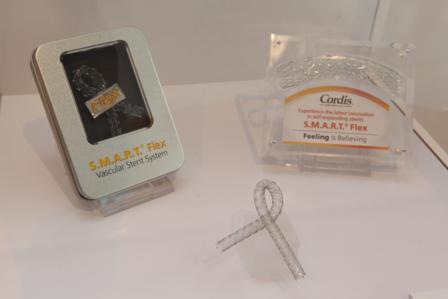
Cordis launched the Smart Flex Self-Expanding Stent System at CX35. The device belongs to a new generation of self-expandable stents for peripheral indications and, according to the company, is “fully connected and yet flexible.”
Laurent Granier, marketing director EMEA, Cardiology and Endovascular at Cordis, spoke to CX Daily News on the features of this new device: “The Smart Flex stent shares a unique design legacy with the Cordis Smart stent—a first generation device. This device does not have any structures that may cause the artery to be injured after some time. The integrity of the stent is expected to be preserved in the long run, meaning that it will be free of fracture, especially in the popliteal and femoral arteries—which are areas where you need this type of stents to be resistant.”
Michael Iwanicky, global marketing leader for Peripheral Stents at Cordis, said that according to the Palmaz principles for stent design, a device should have high radial force and good tissue-metal ratio, and be fully connected. “Cordis has achieved all this with this device; it is fully connected and yet flexible,” Iwanicky said. “The device has been implanted in 200 patients so far”.
Following the recent acquisition of Flexible Stent Solutions—a developer of flexible peripheral arterial, venous and biliary stents—Granier commented, “Cordis has been able to expand the Smart platform to address unmet needs in the treatment of peripheral arterial disease and extend our capabilities to develop therapeutic applications into below-the-knee and venous interventions.”
“If we talk about education, Charing Cross is for us the most important endovascular meeting for vascular surgeons and interventional radiologists in the international European science arena. This is the place where we might effectively have the best impact launching this new generation of the Smart device”. He also commented that the product would be available from June 2013.
In Europe, the device gained the CE mark approval for all peripheral indications (femoral, popliteal and iliac arteries). In the USA, the Smart Flex stent has been approved for biliary indications.
Plain old balloons and bare metal stents do not reign supreme in the superficial femoral artery
After listening to the latest data for the Zilver PTX drug-eluting peripheral stent, 81% of delegates at yesterday’s stent data session voted against the motion that “plain old balloon and bare metal stents reign supreme in the superficial femoral artery”.
At the session, Frank Criado, Baltimore, USA, reviewed whether plain old balloons and bare metal stents were still the first-line treatment devices for the superficial femoral artery. He said that currently, vascular physicians were “addicted to stents” because they are predictable, easy to use, and widely available. According to Criado, there are disadvantages with this approach and these include monetary costs, the risk of in-stent restenosis, fractures and the possible need for re-intervention. However, with current positive data from the RESILIENT trial and the one-year data from the Zilver PTX trial, Criado predicted that the use of plain old balloons and bare metal stents—in the next five years—will be replaced by drug-eluting stents, drug-eluting balloons, mechanical and laser atherectomy, and endoluminal bypass.
In a following presentation, Marc Bosiers (Dendermonde, Belgium) discussed the use of the Zilver PTX in long lesions in the superficial femoral artery (>15cm). He said that stent integrity decreases as lesion length increases, but added that the Zilver PTX “provides better results in long lesions than bare metal stents”. Concluding his presentation, Bosiers announced the launch of the Zilverpass (The Cook Zilver PTX drug-eluting stent versus bypass surgery for the treatment of femoropopliteal TASC C and D lesions) study, which has a primary endpoint of patency at 12 months.
Michael Dake (Stanford, USA) then presented the three-year data from the Zilver PTX (Cook Medical) drug-eluting stent clinical trial. The Zilver PTX, according to Dake, is designed for the superficial femoral artery and is approved in Europe, Japan, and the USA. He reported that the Zilver PTX trial had a primary and secondary randomisation process. According to the results, event-free survival was 83.5% in the 185 patients who received the Zilver PTX stent compared with 72.7% in 189 patients who were treated with percutaneous transluminal angioplasty alone (p<0.01). The rate of freedom from target lesion revascularisation was 84% in the 185 patients and 70.2% in the 189 patients. Dake added that, at three years, the Zilver PTX demonstrated a low fracture rate of 2.1%.
Additionally, the primary patency of the Zilver PTX was 68.7% in 184 lesions vs. 22.8% in 207 lesions with percutaneous transluminal angiography. In a comparison between the Zilver PTX stent and a bare metal stent, the primary patency was 79.6% in 44 lesions and 56.3% in 53 lesions respectively. “Three-year results support sustained safety and effectiveness,” He concluded.
Substantial improvement in functional status with bioresorbable scaffold at 30 days
Yesterday, at the Abbott Vascular Satellite Symposium, Johannes Lammer (Vienna, Austria) presented the 30-day data for a drug-eluting bioresorbable scaffold (Espirit; Abbott Vascular) for the management of de novo lesions in the superficial femoral or iliac arteries—the data showed that the scaffold was associated with a 100% procedural success rate and a substantial improvement in functional status.
Lammer reported that the scaffold was made from poly L-lactide, was naturally resorbed and fully metabolised, and was a hybrid between a balloon-expandable and a self-expanding stent. He added that ESPRIT I study is a single-arm, multicentre and its aim is evaluate the use of the bioresorbable scaffold in patients with a single de novo lesion in the superficial femoral or iliac arteries and who had symptomatic claudication. The trial objectives, Lammer reported, were to evaluate the safety and performance of the scaffold and the endpoints included the procedural, clinical and functional outcomes at one, six, 12 months and at two and three years.
At 30 days, in 35 patients (recruited across seven centres), there were no deaths, no scaffold thrombosis, and no target lesion revascularisations. Also, there was a 100% acute procedural success rate. Furthermore, there was a substantial improvement in functional status—Lammer commented that the percentage of severe claudicants (Rutherford category 3) dropped from 57% at baseline to 0% at 30 days. Concluding his presentation, Lammer said: “So, you can see the initial results are remarkable.”
Delegates at the Satellite Symposium also listened to Frank Vermassen, Ghent, Belgium, speaking about femoral anatomy, characteristics and challenges, and Richard Rapoza, San Francisco, USA, on evolution in superficial femoral artery technology.
Latin America comes to CX
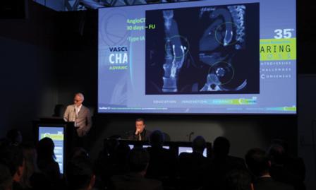
For the first time, as part of the new session “CX Meets Latin America”, Latin American physicians shared their experiences of treating abdominal aortic and thoraco-aortic aneurysm with CX delegates.
At the session entitled “Pushing the boundaries of EVAR and TEVAR”, CX delegates received information on case reports from Argentina, Brazil and Puerto Rico. Frank Criado, Baltimore, USA, chaired the session with Frans Moll, Utretcht, The Netherlands, and Tulio Navarro, Belo Horizonte, Brazil, as moderators.
Mariano Ferreira, Buenos Aires, Argentina, presented a case of a 74-year-old man with a 7cm thoraco-abdominal aneurysm with distal dilatation of the aortic arch that was treated in a three-staged thoracic endovascular aortic repair (TEVAR) procedure. Ferreira said: “Due to elevated spinal cord ischaemic risk, a three-staged TEVAR procedure was planned with prophylactic left subclavian artery revascularisation.”
He explained that the patient underwent TEVAR under general anaesthetic with adjunctive spinal fluid drainage. “Through a direct right femoral access, three Gore Tag thoracic endoprostheses (40 x 140mm, 37 x 200mm and 34 x 200mm) were implanted in an overlapping proximal-to-distal fashion covering the entire length of the thoraco-aortic aneurysm (from the left common carotid artery origin to the celiac axis trunk).”
Ferreira commented that the patient did not present any complications after the procedure and was discharged home on the fourth postoperative day. He added: “Twelve days later, the patient underwent successful percutaneous proximal left subclavian plug embolization to guarantee complete sealing of the thoraco-abdominal aneurysm.” After one-year follow-up, Ferreira concluded, a CT scan revealed complete exclusion of the thoraco-abdominal aneurysm.
A case of a “Hybrid treatment of ascending aorta and arch without opening the chest wall” was presented by Tulio Navarro, Belo Horizonte, Brazil. He said: “The treatment of the aortic arch is one of the greatest challenges to the surgeon. The conventional open repair requires sternotomy and cardiopulmonary bypass. In order to reduce complications, the use of endovascular techniques has been shown to be feasible.”
For this case, Navarro described an alternative approach to ascending aorta, arch and descending aorta with off-the-shelf devices without opening the chest wall. He said: “A 71-year-old male underwent a right-to-left carotid-carotid bypass graft. Using this access, a 16x16x95mm Medtronic limb extension was placed at the ascending aorta, and then a 44x44x200mm TEVAR Valiant-Captivia endograft was also placed at the ascending aorta distal to the limb extension, via right femoral artery.” According to Navarro, the patient tolerated the procedure well and was discharged after three days, asymptomatic and with no stroke. He told the audience that after 17 months of follow-up, the patient was still alive and remained asymptomatic, with no sequels, no strokes and that all grafts were patent.
Navarro also presented a second case, which was of a 58-year-old female with prior type A dissection and who was operated on five years ago. He explained that she presented with hoarseness and chest discomfort. According to Navarro, the CT scan showed an 8cm aortic arch aneurysm affecting all supraortic vessels and chronic dissection going down below visceral aorta. She was treated the same way as the previous case. After four months of follow-up, Navarro commented, there was still remaining hoarseness but no chest discomfort and CT scan did not show any leaks. This technique, Navarro concluded: “allows the treatment of aortic arch diseases without opening the chest wall with off-the-shelf devices with good results.”
However, this alternative approach should only be performed only in high-risk patients and needs to be evaluated with long-term results and larger series.
Also, at the session, several challenging cases from Brazil were presented—endovascular treatment of complex type IV aneurysm by Pierre Galvagni Silveira, Florianopolis, Brazil; a not suitable neck abdominal aortic aneurysm treated by physician fenestrated endograft by Gustavo Puludetto, Brasilia, Brazil; an endovascular treatment of aortic pseudoaneurysm by Alvaro Razuk, Sao Paulo, Brazil; and a case of ascending aorta treated by a heart and vascular team (Eduardo Saadi, Porto Alegre , Brazil).
Criado closed the session saying: “This is a good beginning of CX Meets Latin America; we hope that this will become a feature of CX making it better and bigger with more room for participants and more speakers from other Latin American countries.”