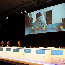
For the third time in CX Symposium history the London-based audience was transported via live video feed to Germany for real-time case presentations. This year CX connected with Arne Schwindt and Theodosios Bisdas in Münster, Germany, for four live peripheral cases alternated with edited cases.
The first live case was performed by Bisdas, who demonstrated spot stenting with VascuFlex Multi-LOC stents combined with the SeQuent Please OTW drug-coated balloon (both from B Braun) in the superior femoral artery (SFA). The team treated a 77-year-old woman presenting with claudication of the right side with Rutherford class 3. The patient had high grade stenosis along the entire SFA.
Bisdas used a transfemoral approach from the left side with a 6F crossover destination sheath followed by plain balloon angioplasty and then a 5mm drug-coated balloon.
Commenting on his reasons for using the VascuFlex Multi-LOC stents, Bisdas said: “We are using it in case we have a flow-limiting dissection that is located at very small lesions along the SFA. In that case I think it is better to use spot stenting so we do not cover the whole SFA. The second point is that, because of recoil after the drug-coated balloon, it is better to use spot stenting to open the lesion than to use a stent of 40mm diameter. The third reason for using this stent is in case of lesions at the popliteal artery where tension might cause stent fracture then I think it is the best option if you have lesions that can be expanded by the radial force of the stent. If you have recoil at the level of the popliteal artery and you do not need to use any atherectomy, then this device is a good option.”
The second live case in the CX Peripheral Live and Edited Cases session was demonstrated by Schwindt and featured a combination technique of atherectomy with the HawkOne device followed by the IN.PACT drug-coated balloon (both from Medtronic) for the treatment of femoropopliteal lesions. He was able to achieve a good outcome with two passes of the atherectomy device—demonstrating the cleaning of the device between passes for the benefit of the audience—and the deployment of the drug-coated balloon.
The third live case, performed again by Schwindt, showed the use of the Supera stent (Abbott Vascular) in highly complex SFA lesions.
The final live case of the day was that of the treatment of an occluded SFA with a Zilver PTX drug-eluting stent (Cook Medical) performed by Bisdas. The patient was a 66-year-old male whose main symptom was claudication with Rutherford class 3. The device was deployed via femoral artery access.
The team explained that the Zilver PTX is a self-expanding drug-eluting stent with paclitaxel. The device, they reported, has been used in over 3,000 post-market cases which have shown notable freedom from target lesion revascularisation.
Much of the discussion surrounding this case was on the sizing of the stent that should be used. After consultation with the moderators and the audience, Bisdas chose to do primary stenting with a 6mm diameter stent, which was only slight oversizing.
The second feature of yesterday’s Peripheral session was the edited cases which were sourced from across the world with presenters from New Zealand, Australia, Belgium, Italy and the USA.
In terms of drug-coated balloons, the audience was treated to edited cases featuring the IN.PACT drug-coated balloon (Medtronic). Andrew Holden (Auckland, New Zealand) showed a case of the use of a drug-coated balloon in the treatment of arteriovenous (AV) fistula. In discussion with the audience, Holden gave his personal tips for a successful procedure.
“The most important procedural step in AV access circuit drug-coated balloon angioplasty is adequate pre-dilatation of stenoses to nominal diameter when compared to the adjacent normal vessel diameter. If there is post-stenotic dilatation of the vein distal to the stenosis, the balloon should be sized to the closest segment of normal calibre vein beyond the dilated segment. At the AV anastomosis, the balloon should be sized to or just above the diameter of the artery,” he explained.
Another highlight of the edited cases was the demonstration of the Tigris stent (Gore) used with intravascular ultrasound (IVUS). Ian Spark (Adelaide, Australia) showed his case of a 72-year-old male patient with rest pain in his left foot and shin ulceration. Preoperative computed tomography showed extensive calcification in the superior femoral artery.
Spark reported that the difference between IVUS and angiography was clearly seen. He said “angiography produces a ‘lumenogram’ with virtually no information about the vessel wall except what can be implied from the shape of the lumen and presence of calcium. IVUS images the entire vessel so that in addition to the lumen the anatomical and pathological details of the wall can be clearly demonstrated.”
The Tigris stent offers a hybrid design with nitinol wire rings and heparin-bonded interconnecting ePTFE lattice. Conventional nitinol stents, Spark noted, are often prone to kinking, fracture and in-stent restenosis.
Commenting on the Tigris stent, Iris Baumgartner (Bern, Switzerland) said: “The Tigris is very flexible. It also mimics the flexibility of the vessel in this critical region and you can do the post-dilatation which is not possible with other stents.”
The remaining edited cases featured the Legflow drug-coated balloon (Cardionovum) in a challenging lesion and the Viabahn endoprosthesis (Gore) in the treatment of in-stent restenosis.