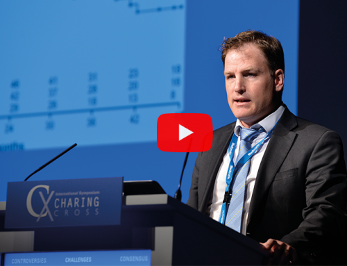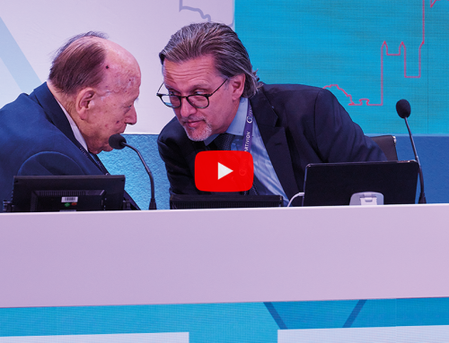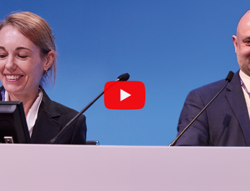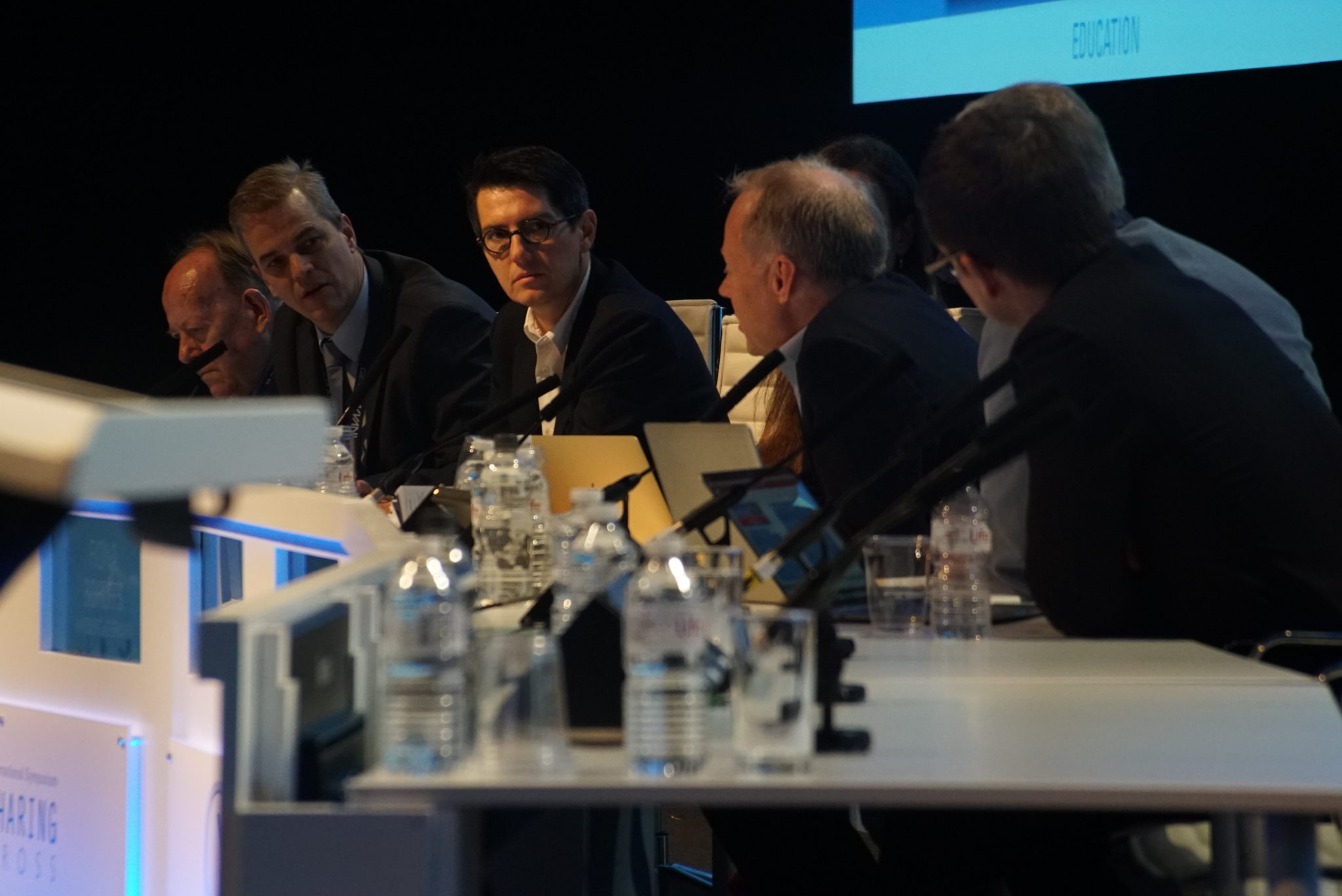
Novel data from the Stroke from Thoracic Endovascular Repair (STEP) collaborators has provided insight into current practice of thoracic endovascular aortic repair (TEVAR), and the future role of diffusion-weighted magnetic resonance imaging (DW-MRI) in reducing stroke from endovascular repair. The multicentre dataset was presented at a Charing Cross (CX) Highlight Session, chaired by study investigator Roger Greenhalgh, and featuring a panel of moderators involved with the study—including Tilo Kölbel (Hamburg, Germany), Stéphan Haulon (Le Plessis Robinson, France), Fiona Rohlffs (Hamburg, Germany), Hugh Markus (Cambridge, UK), Heinz Jakob (Essen, Germany) and Gustavo Oderich (Rochester, USA).
Taking a collaborative approach to investigating stroke risks associated with thoracic aortic repair, STEP is an independent, all-encompassing, open and interdisciplinary study aiming to provide best practice for endovascular procedures, ultimately lowering the risk of cerebral embolism.
The STEP collaborators have reported that “currently, better evaluation is needed” in terms of cerebral infarction and cognitive function after performing thoracic endovascular procedures. “There should be a better way to quantify what happens to the brain from cerebral infarction, embolism, haemorrhage and even thrombus,” Rohlffs said in an introduction of the session at CX. For this reason, she explained, the STEP collaborators worked to pool experience from participating centres in the USA and Europe.
Summarising the major conclusions presented last year at CX, Rohlffs stated that with the pooled experience and multidisciplinary guidance from specialists in neurology, interventional cardiology, cardiothoracic surgery and neuroradiology, “we learned that imaging is a crucial tool for us to evaluate stroke after the procedure, as well as silent brain lesions that can occur after TEVAR.” The main aim of STEP, noted Greenhalgh, is to investigate “what infarctions there are, the type of infarctions after the procedure, and whether surgical technique can improve.” It was highlighted that, as surgeons trying to recognise clinical stroke may underdiagnose this condition, the potential value of introducing DW-MRI was evaluated in phase 2 of the STEP collaboration.
This potential value, the panel explained, lies in the ability of DW-MRI to recognise whether a lesion appearing in follow-up imaging is associated with the procedure itself, while also providing a means to monitor the outcome and improvement in the long-term. From the panel, Markus noted that what is “really exciting” about DW MRI in this area, is clear “when looking at it for improving surgical endovascular technique”.
Among the STEP operators, three centres (Hamburg, Germany; Paris, France and the MayoClinic, USA) were able to provide new data on the use of DW-MRI post-TEVAR procedures, involving zone 0, 1, 2 and 3 of the aortic arch. The total series of 37 cases combined from these centres presents the “largest global experience” to date, Greenhalgh noted.
According to Kölbel, “the biggest difference to what is already known is that these cases include a large number of very proximal landings—with a significant zone 0 and zone 1 percentage. It is important that MRI is done within a certain post-procedural time frame, and we were able to keep to the range of two to eight days, with the exception of only one patient.” He added, this suggested time frame of up to eight days was established with advice from Markus. Haulon expanded on the finding that a high proportion of cases were performed in the most proximal landing zones: “what was interesting about this dataset was that in less than three months, we managed to gather over 20 cases with proximal landing in zone 0 and 1, equating to almost 60% of the entire cohort.”
A second unanticipated result was the key finding that while new lesions were seen on DW-MRI in 80% of zone 0 cases, “63% of zone 3 patients were found to have new lesions on DW-MRI”, Haulon highlighted. These new lesions were seen despite the investigators use of preventative measures, including carotid clamping and, notably, CO2 flushing of the device systems before the standard saline flush. The latter technique was introduced by Kölbel and Rohlffs, whose work suggests the CO2 flush of stent grafts may help reduce air embolisation during TEVAR.
However, through a survey of collaborators regarding current practice, the limitation for DW-MRI funding was brought to light, as the majority of centres were unable to make use of the imaging modality. “We found that in the US centres where STEP collaborators operate, they cannot get funding for DW-MRI,” Greenhalgh explained. “However, perhaps the institutions could be persuaded, that if they perform this scan in the post-operative period, it can be recognised whether infarcts seen on imaging were present prior to the procedure.” As a result, an argument for funding could be made, as centres could distinguish between procedurally associated harm and pre-existing lesions.
Nevertheless, Markus warned that despite the “interesting findings” of the study and potential for DW-MRI to improve endovascular practice, “it remains important to have cognitive testing, to determine the impact thoracic endovascular repair may have on the patient.” This was elaborated on by Greenhalgh, who pointed to the fact that “some cognitive symptoms of neural damage may appear, even without visible DW-MRI lesions.”
Both Markus and Greenhalgh remarked that future investigation will require a larger number of patients—both for improved understanding of the value of DW-MRI and current TEVAR practice, as well as to ensure adequate power in the analysis of cognitive testing data.
Concluding the session, Greenhalgh argued: “DW-MRI should be a standard of care—by which I mean that whether open or endovascular, we should press upon the funding bodies of our hospitals and institutions, that the procedures should include funded DW-MRI.”


