Mark Whiteley (London, UK), member of the CX Programme Organising Board, discusses various techniques for superficial venous disease treatment and their applications to specific patients. The latest evidence on this area will be discussed at the CX Venous Challenges Day (26 April 2016).
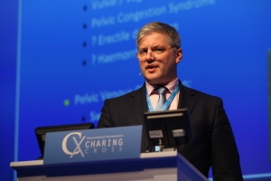
Mark Whiteley
Could you please give us an overview of the various techniques and applications for superficial venous treatment?
I believe that endovenous thermal ablation is still the gold standard treatment for endovenous ablation. If endovenous thermoablation is performed correctly, using the best principles, failure of closure or late recanalisation should be a rarity.
However, thermal ablation cannot be used in some specific areas such as in truncal veins under hostile tissue, truncal veins next to important nerves or even in pelvic veins, where thermal spread might damage adjacent organs or structures. As such, alternative non-thermal techniques should be viewed both in terms of use in these specific indications as well as being in competition with thermal techniques in the truncal veins of the legs.
In view of our recently published results of the effect of sclerotherapy in vein walls, foam sclerotherapy alone should be reserved for the smaller or thin walled veins, where it can have an excellent result. There are adjuvant techniques that can be used to help improve the efficacy of foam sclerotherapy such as tumescence. It may well be that researchers in the field can combine such adjuvant techniques to improve foam sclerotherapy sufficiently to challenge thermal ablation in thick walled truncal veins.
Additionally, I consider that mechanochemical ablation (MOCA) has many advantages over thermal ablation, and is ideal to be used in veins next to nerves or under hostile tissue. As a treatment for truncal veins, it avoids the need for injections of tumescence, but can be limited by sclerotherapy dose. However, there are newer strategies that are improving this.
What could you say regarding the use of cyanoacrylate adhesive embolisation?
Cyanoacrylate glue is a fascinating development, which has shown that there are different ways to cause ablation. Whilst all thermal and chemical ablations rely on cellular damage through apoptosis to cause fibrosis, cyanoacrylate glue works by stimulating a foreign body reaction. This has been shown to work, resulting in fibrosis and is probably the same reaction as that caused by embolisation coils.
However, although quick, relatively easy to perform and requiring only one injection per vein, it does have some drawbacks at the current time. Cost is the most quoted. There are some other questions regarding mechanisms of action, the biggest and smallest veins that can be treated, how it can be used in treating incompetent perforating veins, etc. With some good basic research we will find out what role cyanoacrylate might actually have in the future of endovenous intervention.
At the Superficial Venous Challenges session of the CX Venous Challenges Day you will be speaking about a method which helps to determine the optimal choice for endovenous treatment; could you give us a few insights from your talk?
When I first spoke at the Charing Cross Symposium in 2004, I presented my views that the key to successful endovenous thermo-ablation was transmural destruction of the vein wall.
At that time, based on some very early ex-vivo histology and basic understanding of pathology, it was clear to me that damage to the endothelium whilst leaving the media alive and intact would lead to acute thrombosis followed by recanalisation of the vein. This would result in poor long-term results. This has now been observed in many devices or techniques that produce this damage profile. They appear to have high early closure rates only to show recanalisation, which results in recurrent reflux in the medium to long term. As such I distrust any device or technique that claims high early occlusion rates of the target vein, which then start reducing at six months, one, two and five years.
In contrast, transmural death of the vein wall without the wall losing its structure, allows the vein to fibrose leading to permanent closure of the target vein and no recurrence of venous reflux neither recurrent reflux in the treated vein. This is what I would call adequate treatment. Indeed, veins treated this way need not be reported as closed as the vein is usually atrophied by six to 12 months, and therefore can be reported as atrophied or even absent. This is what we found in our 12-year follow-up study which we presented a few years ago at both SITE and at the Venous Forum of the Royal Society of Medicine.
Of course, just as one can undertreat the vein, there is also the risk of overtreatment. In this circumstance, instead of just transmural death of the cells, the vein wall is so damaged that becomes disrupted. This can result in intense pain due to necrotic tissue stimulating an intense inflammatory reaction, or it can result in disruption of the vein wall, allowing localised loss of luminal contents resulting in bruising or even haematoma.
As such, we need to be able to quantify how much total ablation energy is needed and how it should be applied to ensure transmural death of cells without resulting in overtreatment and necrosis in the inner layers of the vein wall.
The CX Venous Challenges Day will take place at the Charing Cross Symposium on Tuesday 26 April – Lower Main Auditorium, Olympia Grand, London, UK
Click here to see the CX Interactive Main Programme – Venous Challenges
Click here to see the CX Venous Workshop
Click here to see the CX Venous Abstract Presentations
Click here to register


